| 存储条件 |
|---|
| Product Name | NF-kB p65/RelA Rabbit mAb |
|---|---|
| Antibody Type | Primary Antibodies |
| Immunogen | A synthetic peptide corresponding to a sequence within amino acids 450-551 of human NF-kB p65/RelA (NP_068810.3). |
| Clonality | monoclonal |
|---|---|
| Isotype | IgG |
| Host Species | Rabbit |
| Tested Applications | ChIPICC/IFIHCIPWB |
| WB:1:10000-1:60000 IHC:1:200-1:2000 ICC/IF:1:100-1:800 IP:0.5μg-4μg antibody for 400μg-600μg extracts of whole cells. ChIP:5μg antibody for 10μg-15μg of Chromatin. | |
| Species Reactivity | HumanMouseRat |
| Concentration | 1mg/ml |
| Purification | Affinity purified |
| Gene Symbol | RELA |
|---|---|
| Gene Synonyms | p65 CMCU NFKB3 AIF3BL3 |
| Gene Full Name | RELA proto-oncogene, NF-kB subunit |
| Gene Summary | NF-kappa-B is a ubiquitous transcription factor involved in several biological processes. It is held in the cytoplasm in an inactive state by specific inhibitors. Upon degradation of the inhibitor, NF-kappa-B moves to the nucleus and activates transcription of specific genes. NF-kappa-B is composed of NFKB1 or NFKB2 bound to either REL, RELA, or RELB. The most abundant form of NF-kappa-B is NFKB1 complexed with the product of this gene, RELA. Four transcript variants encoding different isoforms have been found for this gene. [provided by RefSeq, Sep 2011] |
| Alternative Names | p65 CMCU NFKB3 AIF3BL3 lA |
| Molecular Weight(MW) | 65kDa |
| Cellular Localization | Cytoplasm, Nucleus. |
WB
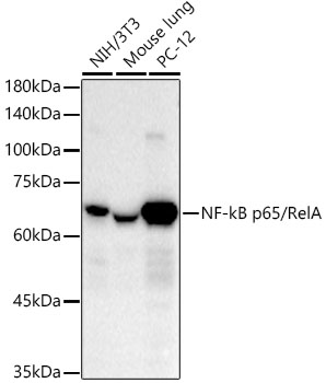
Western blot analysis of various lysates using NF-kB p65/RelA Rabbit mAb at 1:10000 dilution. Secondary antibody: HRP-conjugated Goat anti-Rabbit IgG (H+L) at 1:10000 dilution. Lysates/proteins: 25μg per lane. Blocking buffer: 3% nonfat dry milk in TBST. Detection: ECL Basic Kit. Exposure time: 30s.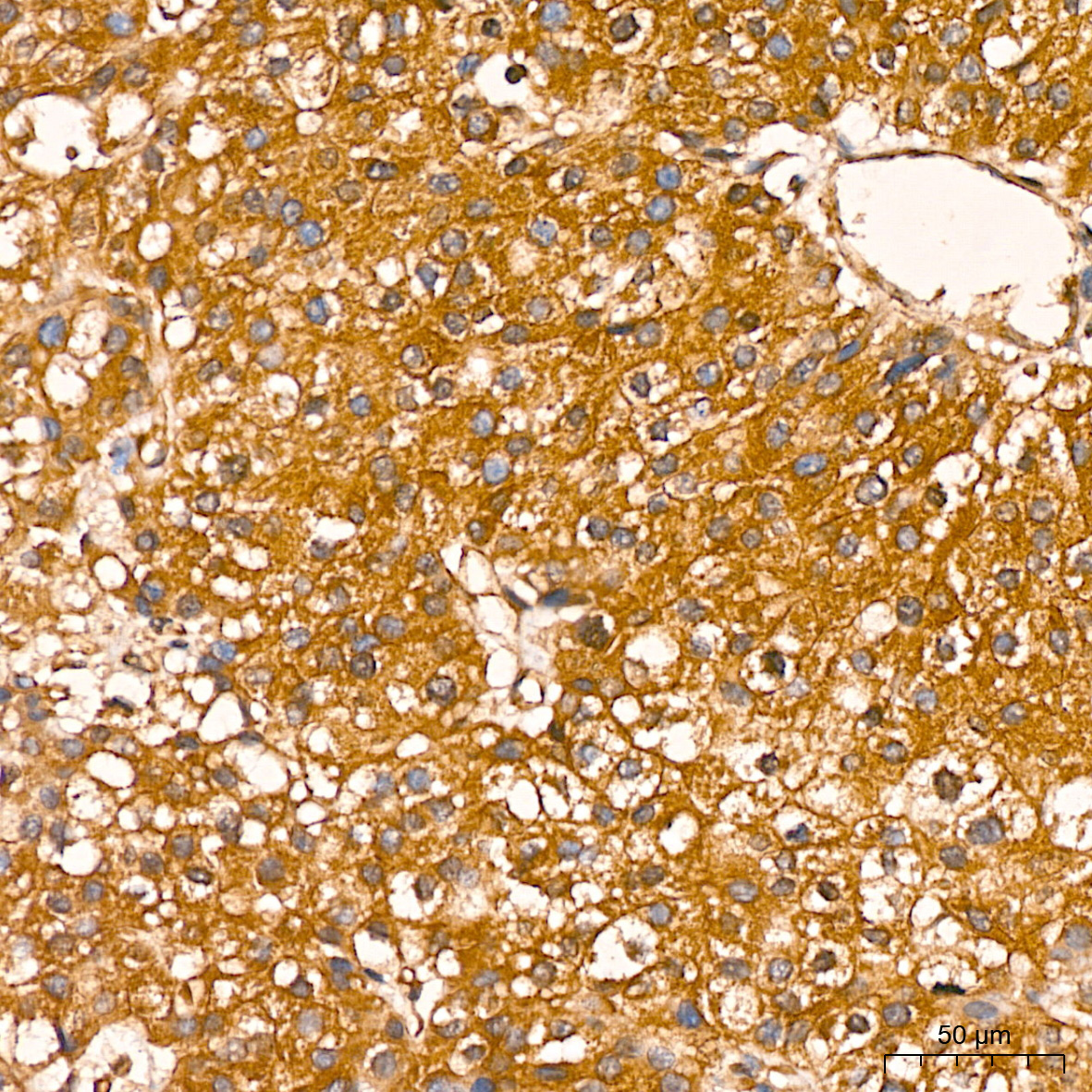
IHC
Immunohistochemistry analysis of paraffin embedded Human liver using NF-kB p65/RelA Rabbit mAb at dilution of 1:200 (40x lens). High pressure antigen retrieval performed with 0.01M Citrate Bufferr (pH 6.0) prior to IHC staining.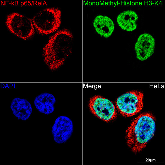
ICC/IF
Confocal imaging of HeLa cells using NF-kB p65/RelA Rabbit mAb (dilution 1:100) (Red). The cells were counterstained with MonoMethyl Histone H3-K4 Rabbit mAb (dilution 1:300) (Green). DAPI was used for nuclear staining (blue). Objective: 60x.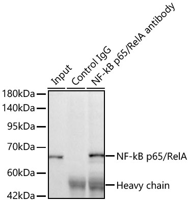
IP
Immunoprecipitation of NF-kB p65/RelA Rabbit mAb from 500 µg extracts of HeLa cells was performed using 2 µg of NF-kB p65/RelA Rabbit mAb. Rabbit IgG isotype control was used to precipitate the Control IgG sample. IP samples were eluted with 1X Laemmli Buffer. The Input lane represents 10% of the total input. Western blot analysis of immunoprecipitates was conducted using NF-kB p65/RelA Rabbit mAb at a dilution of 1:10000.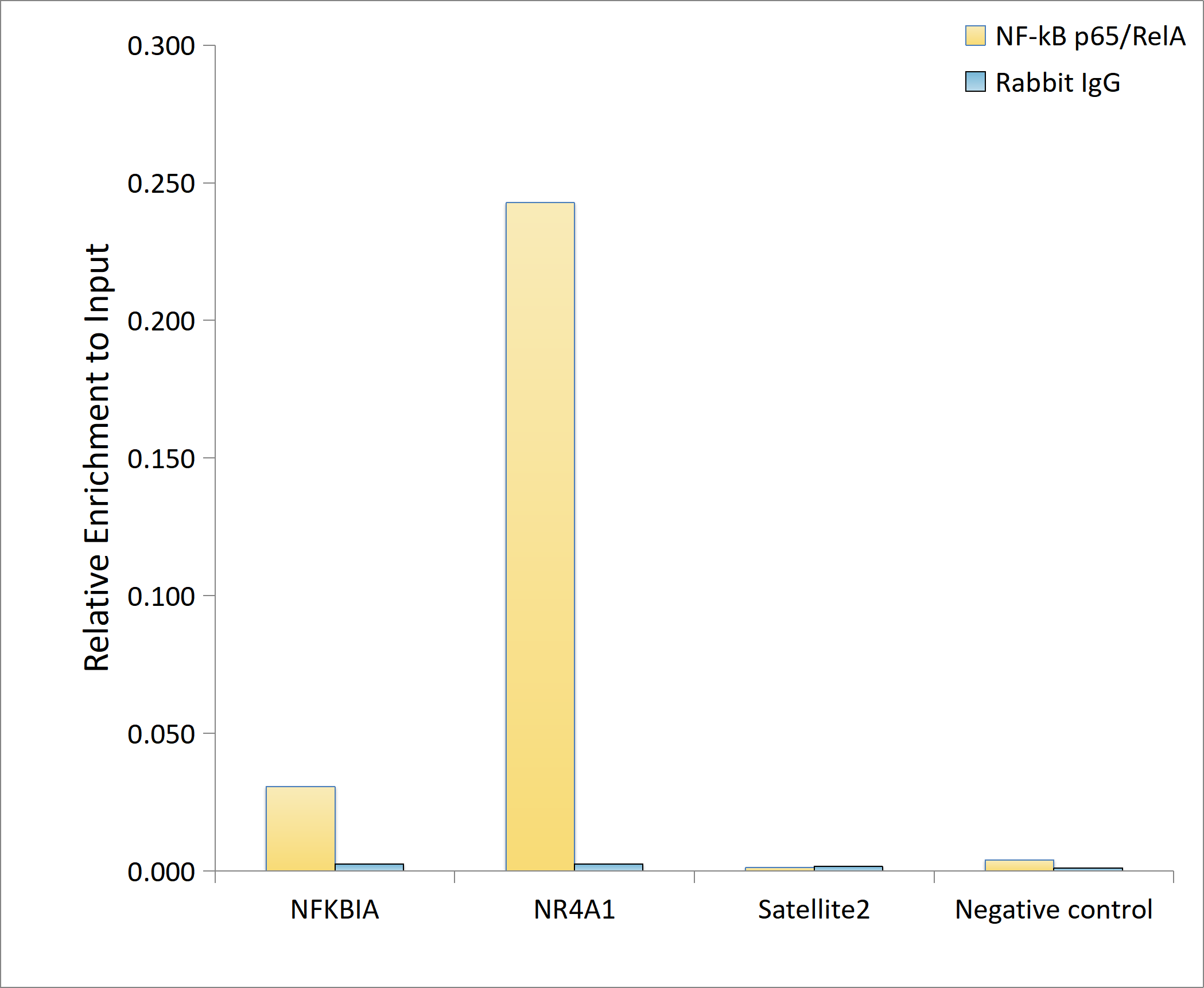
ChIP
Chromatin immunoprecipitation analysis of extracts of HT-1080 cells, HT-1080 cells were treated by TNF-α (20 ng/ml) at 37℃ for 30 minutes, using NF-kB p65/RelA antibody and rabbit IgG.The amount of immunoprecipitated DNA was checked by quantitative PCR. Histogram was constructed by the ratios of the immunoprecipitated DNA to the input.| Application Notes | WB:1:10000-1:60000 IHC:1:200-1:2000 ICC/IF:1:100-1:800 IP:0.5μg-4μg antibody for 400μg-600μg extracts of whole cells. ChIP:5μg antibody for 10μg-15μg of Chromatin. |
|---|
| Form | Liquid |
|---|---|
| Storage Instructions | Store at -20℃. Avoid freeze / thaw cycles. |
| Storage Buffer | Buffer: PBS with 0.05% proclin300, 0.05% BSA, 50% glycerol, pH7.3. |
抱歉,暂无相关文献







