| 存储条件 |
|---|
| Product Name | Annexin A1/ANXA1 Rabbit mAb |
|---|---|
| Antibody Type | Primary Antibodies |
| Immunogen | Recombinant fusion protein containing a sequence corresponding to amino acids 2-346 of human Annexin A1/ANXA1 (NP_000691.1). |
| Clonality | monoclonal |
|---|---|
| Isotype | IgG |
| Host Species | Rabbit |
| Tested Applications | ICC/IFIHCIPWB |
| WB:1:7000-1:70000 IHC:1:2000-1:20000 ICC/IF:1:200-1:400 IP:0.5μg-4μg antibody for 400μg-600μg extracts of whole cells. | |
| Species Reactivity | HumanMouseRat |
| Concentration | 1mg/ml |
| Purification | Affinity purified |
| Gene Symbol | ANXA1 |
|---|---|
| Gene Synonyms | ANX1 LPC1 |
| Gene Full Name | annexin A1 |
| Gene Summary | This gene encodes a membrane-localized protein that binds phospholipids. This protein inhibits phospholipase A2 and has anti-inflammatory activity. Loss of function or expression of this gene has been detected in multiple tumors. [provided by RefSeq, Dec 2014] |
| Alternative Names | ANX1 LPC1 |
| Molecular Weight(MW) | 38kDa |
| Cellular Localization | Apical cell membrane, Basolateral cell membrane, Cell membrane, Cell projection, Cytoplasm, Cytoplasmic vesicle, Cytoplasmic vesicle membrane, Early endosome, Endosome membrane, Extracellular side, Lateral cell membrane, Membrane, Nucleus, Peripheral membrane protein, Secreted, cilium, exosome, extracellular space, phagocytic cup, secretory vesicle lumen. |
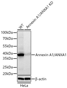
WB
Western blot analysis of lysates from wild type (WT) and Annexin A1/ANXA1 knockdown (KD) HeLa cells using Annexin A1/ANXA1 Rabbit mAb at 1:8000 dilution incubated overnight at 4℃. Secondary antibody: HRP-conjugated Goat anti-Rabbit IgG (H+L)at 1:10000 dilution. Lysates/proteins: 25 μg per lane. Blocking buffer: 3% nonfat dry milk in TBST. Detection: ECL Basic Kit. Exposure time: 1s.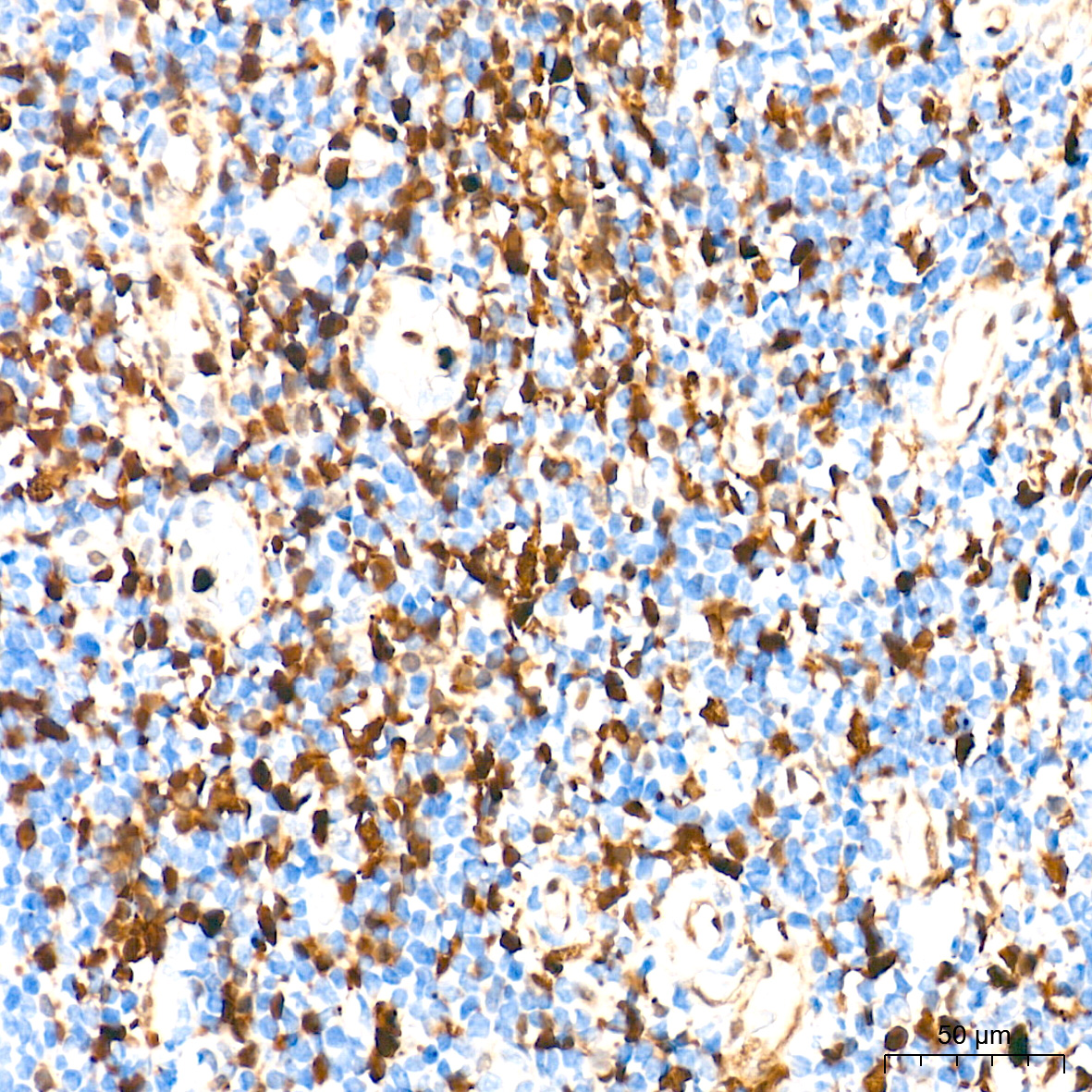
IHC
Immunohistochemistry analysis of paraffin embedded Human tonsil tissue using Annexin A1/ANXA1 Rabbit mAb at a dilution of 1:9000 (40x lens). High pressure antigen retrieval performed with 0.01M Tris-EDTA Buffer(pH 9.0) prior to IHC staining.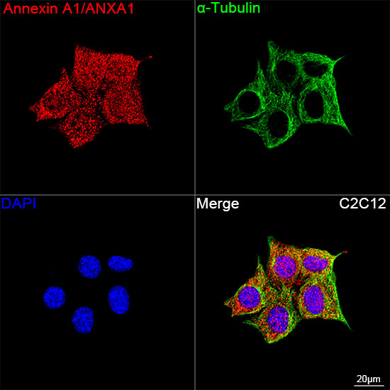
ICC/IF
Confocal imaging of C2C12 cells using Annexin A1/ANXA1 Rabbit mAb (dilution 1:200) followed by a further incubation with Cy3 Goat Anti-Rabbit IgG (H+L)(dilution 1:500) (Red). The cells were counterstained with α-Tubulin Mouse mAb (dilution 1:400) followed by incubation with Omnimabs® 488-conjugated Goat Anti-Mouse IgG (H+L) Ab (dilution 1:500) (Green). DAPI was used for nuclear staining (Blue). Objective: 100x.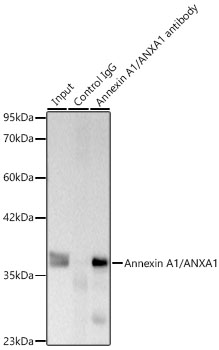
IP
Immunoprecipitation of Annexin A1/ANXA1 from 500 µg extracts of C2C12 cells was performed using 2 µg of Annexin A1/ANXA1 Rabbit mAb. Rabbit IgG isotype control was used to precipitate the Control IgG sample. IP samples were eluted with 1X Laemmli Buffer. The Input lane represents 10% of the total input. Western blot analysis of immunoprecipitates was conducted using Annexin A1/ANXA1 Rabbit mAb at a dilution of 1:8000.| Application Notes | WB:1:7000-1:70000 IHC:1:2000-1:20000 ICC/IF:1:200-1:400 IP:0.5μg-4μg antibody for 400μg-600μg extracts of whole cells. |
|---|
| Form | Liquid |
|---|---|
| Storage Instructions | Store at -20℃. Avoid freeze / thaw cycles. |
| Storage Buffer | Buffer: PBS with 0.05% proclin300, 0.05% BSA, 50% glycerol, pH7.3. |
抱歉,暂无相关文献







