| 存储条件 |
|---|
| Product Name | KPNB1 Recombinant Rabbit Monoclonal Antibody |
|---|---|
| Antibody Type | Primary Antibodies |
| Immunogen | Recombinant protein within human KPNB1 aa 1-550. |
| Clonality | monoclonal |
|---|---|
| Isotype | IgG |
| Host Species | Rabbit |
| Tested Applications | IF-PIHCIPWB |
| WB:1:1000 IHC:1:1000 IF-P:1:200 IP:1-2μg/sample | |
| Species Reactivity | HumanMouseRat |
| Concentration | 1mg/ml |
| Purification | Protein A |
| Gene Symbol | KPNB1 |
|---|---|
| Gene Synonyms | IMB1 IPO1 IPOB Impnb NTF97 |
| Gene Full Name | karyopherin subunit beta 1 |
| Gene Summary | Nucleocytoplasmic transport, a signal- and energy-dependent process, takes place through nuclear pore complexes embedded in the nuclear envelope. The import of proteins containing a nuclear localization signal (NLS) requires the NLS import receptor, a heterodimer of importin alpha and beta subunits also known as karyopherins. Importin alpha binds the NLS-containing cargo in the cytoplasm and importin beta docks the complex at the cytoplasmic side of the nuclear pore complex. In the presence of nucleoside triphosphates and the small GTP binding protein Ran, the complex moves into the nuclear pore complex and the importin subunits dissociate. Importin alpha enters the nucleoplasm with its passenger protein and importin beta remains at the pore. Interactions between importin beta and the FG repeats of nucleoporins are essential in translocation through the pore complex. The protein encoded by this gene is a member of the importin beta family. Two transcript variants encoding different isoforms have been found for this gene. [provided by RefSeq, Feb 2013] |
| Alternative Names | IMB 1 antibody IMB1 antibody IMB1_HUMAN antibody Impnb antibody Importin 90 antibody Importin beta 1 antibody Importin beta 1 subunit antibody Importin subunit beta-1 antibody Importin-90 antibody IPOB antibody IMB 1 antibody IMB1 antibody IMB1_HUMAN antibody Impnb antibody Importin 90 antibody Importin beta 1 antibody Importin beta 1 subunit antibody Importin subunit beta-1 antibody Importin-90 antibody IPOB antibody Karyopherin beta 1 antibody Karyopherin beta 1 subunit antibody Karyopherin subunit beta-1 antibody KPNB 1 antibody Kpnb1 antibody MGC2155 antibody MGC2156 antibody MGC2157 antibody NTF 97 antibody NTF97 antibody NTF97/Importin beta antibody Nuclear factor P97 antibody Pore targeting complex 97 kDa subunit antibody PTAC97 antibody |
| Molecular Weight(MW) | 97kDa |
| Cellular Localization | Cytoplasm. Nucleus envelope. |
WB
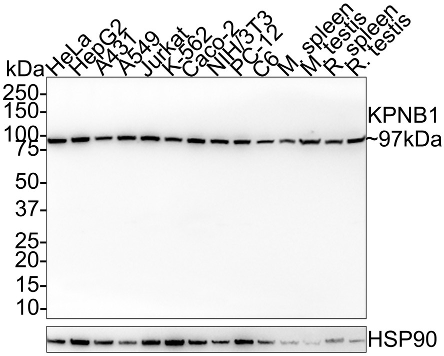
Western blot analysis of KPNB1 on different lysates with Rabbit anti-KPNB1 antibody at 1/1,000 dilution. Lane 1: HeLa cell lysate (20 µg/Lane), Lane 2: HepG2 cell lysate (20 µg/Lane), Lane 3: A431 cell lysate (20 µg/Lane), Lane 4: A549 cell lysate (20 µg/Lane), Lane 5: Jurkat cell lysate (20 µg/Lane), Lane 6: K-562 cell lysate (20 µg/Lane), Lane 7: Caco-2 cell lysate (20 µg/Lane), Lane 8: NIH/3T3 cell lysate (20 µg/Lane), Lane 9: PC-12 cell lysate (20 µg/Lane), Lane 10: C6 cell lysate (20 µg/Lane), Lane 11: Mouse spleen tissue lysate (40 µg/Lane), Lane 12: Mouse testis tissue lysate (40 µg/Lane), Lane 13: Rat spleen tissue lysate (40 µg/Lane), Lane 14: Rat testis tissue lysate (40 µg/Lane), Exposure time: 1 minute; 4-20% SDS-PAGE gel. Proteins were transferred to a PVDF membrane and blocked with 5% NFDM/TBST for 1 hour at room temperature. The primary antibody at 1/1,000 dilution was used in 5% NFDM/TBST at room temperature for 2 hours. Goat Anti-Rabbit IgG - HRP Secondary Antibody at 1/100,000 dilution was used for 1 hour at room temperature.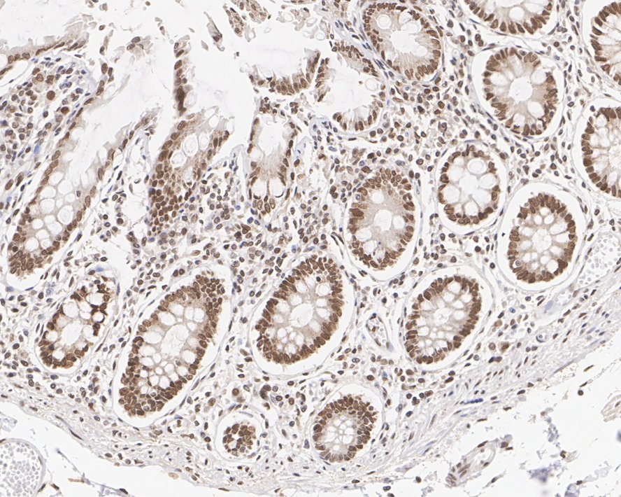
IHC
Immunohistochemical analysis of paraffin-embedded human colon tissue with Rabbit anti-KPNB1 antibody at 1/1,000 dilution. The section was pre-treated using heat mediated antigen retrieval with sodium citrate buffer (pH 6.0) for 2 minutes. The tissues were blocked in 1% BSA for 20 minutes at room temperature, washed with ddH2O and PBS, and then probed with the primary antibody at 1/1,000 dilution for 1 hour at room temperature. The detection was performed using an HRP conjugated compact polymer system. DAB was used as the chromogen. Tissues were counterstained with hematoxylin and mounted with DPX.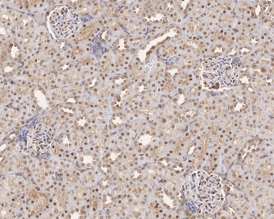
IHC
Immunohistochemical analysis of paraffin-embedded rat kidney tissue with Rabbit anti-KPNB1 antibody at 1/1,000 dilution. The section was pre-treated using heat mediated antigen retrieval with sodium citrate buffer (pH 6.0) for 2 minutes. The tissues were blocked in 1% BSA for 20 minutes at room temperature, washed with ddH2O and PBS, and then probed with the primary antibody at 1/1,000 dilution for 1 hour at room temperature. The detection was performed using an HRP conjugated compact polymer system. DAB was used as the chromogen. Tissues were counterstained with hematoxylin and mounted with DPX.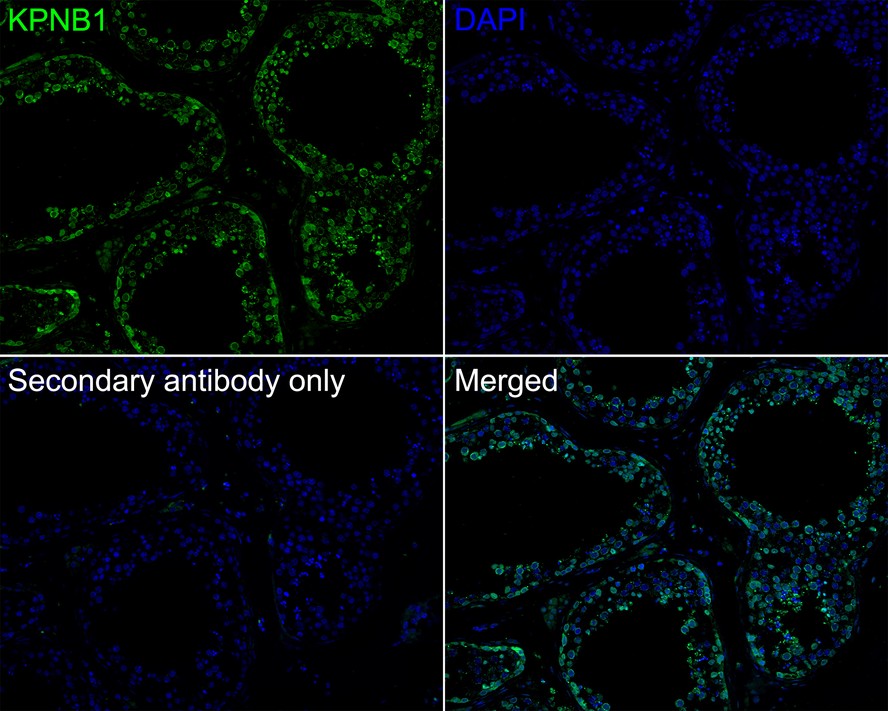
IF-P
Immunofluorescence analysis of paraffin-embedded human testis tissue labeling KPNB1 with Rabbit anti-KPNB1 antibody at 1/200 dilution. The section was pre-treated using heat mediated antigen retrieval with Tris-EDTA buffer (pH 9.0) for 20 minutes. The tissues were blocked in 10% negative goat serum for 1 hour at room temperature, washed with PBS, and then probed with the primary antibody ( green) at 1/200 dilution overnight at 4 ℃, washed with PBS. Goat Anti-Rabbit IgG H&L (488) was used as the secondary antibody at 1/1,000 dilution. Nuclei were counterstained with DAPI (blue).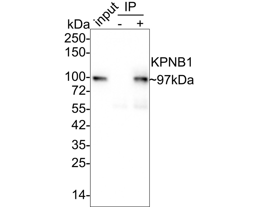
IP
KPNB1 was immunoprecipitated in 0.2mg HeLa cell lysate with Rabbit anti-KPNB1 antibody at 2 µg/25 µl agarose. Western blot was performed from the immunoprecipitate using Rabbit anti-KPNB1 antibody at 1/1,000 dilution. Anti-Rabbit IgG for IP Nano-secondary antibody at 1/5,000 dilution was used for 1 hour at room temperature. Lane 1: HeLa cell lysate (input), Lane 2: Rabbit IgG instead of Rabbit anti-KPNB1 antibody in HeLa cell lysate, Lane 3:Rabbit anti-KPNB1 antibody IP in HeLa cell lysate, Blocking/Dilution buffer: 5% NFDM/TBST, Exposure time: 24 seconds.| Application Notes | WB:1:1000 IHC:1:1000 IF-P:1:200 IP:1-2μg/sample |
|---|
| Form | Liquid |
|---|---|
| Storage Instructions | Store at +4℃ after thawing. Aliquot store at -20℃. Avoid repeated freeze / thaw cycles. |
| Storage Buffer | PBS (pH7.4), 0.1% BSA, 40% Glycerol. Preservative: 0.05% Sodium Azide. |
抱歉,暂无相关文献







