| 存储条件 |
|---|
| Product Name | Rabbit polyclonal antibody to Filaggrin |
|---|---|
| Antibody Type | Primary Antibodies |
| Immunogen | A synthesized peptide derived from human FLG(Accession P20930), corresponding to amino acid residues E3961-R4011. |
| Clonality | Polyclonal |
|---|---|
| Isotype | IgG |
| Host Species | Rabbit |
| Tested Applications | ICC/IFIHCWB |
| WB:1:500-1:2000 IHC:1:50-1:200 ICC/IF:1:100-1:500 | |
| Species Reactivity | HumanRat |
| Concentration | 1mg/ml |
| Purification | Affinity purified |
| Gene Symbol | FLG |
|---|---|
| Gene Synonyms | FLG1 ATOD2 FLG-1 |
| Gene Full Name | filaggrin |
| Gene Summary | The protein encoded by this gene is an intermediate filament-associated protein that aggregates keratin intermediate filaments in mammalian epidermis. It is initially synthesized as a polyprotein precursor, profilaggrin (consisting of multiple filaggrin units of 324 aa each), which is localized in keratohyalin granules, and is subsequently proteolytically processed into individual functional filaggrin molecules. Mutations in this gene are associated with ichthyosis vulgaris.[provided by RefSeq, Dec 2009] |
| Alternative Names | ATOD2 Epidermal filaggrin FILA_HUMAN Filaggrin Filaggrin precursor Fillagrin FLG Profilaggrin |
| Molecular Weight(MW) | 30-120kDa; 435kDa(Calculated). |
| Cellular Localization | Cytoplasmic granule. |
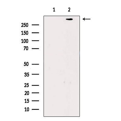
WB
Western blot analysis of extracts from Rat eyes, using Filaggrin Antibody. The lane on the left was treated with blocking peptide.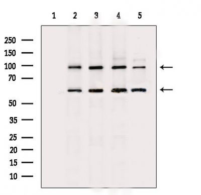
WB
Western blot analysis of extracts from various samples, using Filaggrin Antibody. Lane 1: K562 cells, blocked with antigen-specific peptides, Lane 2: K562 cells, Lane 3: HepG2 cells, Lane 4: A2789 cells, Lane 5: MDA-MB-231 cells.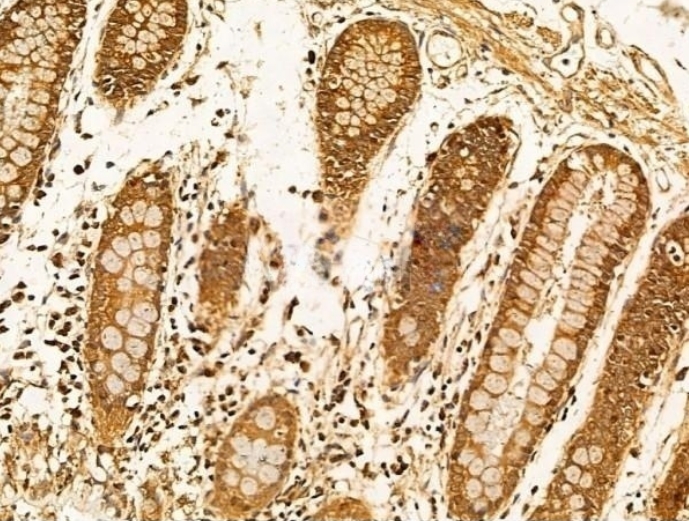
IHC
Filaggrin Antibody at 1/100 staining Human esophageal cancer by IHC-P. The sample was formaldehyde fixed and a heat mediated antigen retrieval step in citrate buffer was performed. The sample was then blocked and incubated with the primary antibody at 4°C overnight. An HRP conjugated anti-Rabbit antibody was used as the secondary antibody.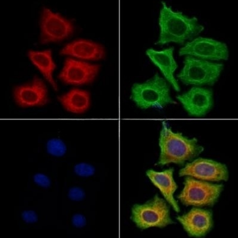
ICC/IF
Filaggrin Antibody staining Hela cells by ICC/IF. The samples were fixed with PFA and permeabilized in 0.1% Triton X-100,then blocked in 10% serum for 45 minutes at 25°C. Samples were then incubated with primary Ab(1:200) and mouse anti-beta tubulin Ab(1:200) for 1 hour at 37°C. An AlexaFluor594 conjugated goat anti-rabbit IgG(H+L) Ab(Red) and an AlexaFluor488 conjugated goat anti-mouse IgG(H+L) Ab(Green) were used as the secondary antibody. The nuclear counter stain is DAPI(blue).| Application Notes | WB:1:500-1:2000 IHC:1:50-1:200 ICC/IF:1:100-1:500 |
|---|
| Form | Liquid |
|---|---|
| Storage Instructions | Store at -20 °C. Stable for 12 months from date of receipt. |
| Storage Buffer | Rabbit IgG in phosphate buffered saline , pH 7.4, 150mM NaCl, 0.02% sodium azide and 50% glycerol. |
抱歉,暂无相关文献







