| 存储条件 |
|---|
| Product Name | SLP76 Recombinant Rabbit Monoclonal Antibody |
|---|---|
| Antibody Type | Primary Antibodies |
| Immunogen | Synthetic phospho-peptide corresponding to residues surrounding Tyr701 of Human STAT1. |
| Clonality | monoclonal |
|---|---|
| Isotype | IgG |
| Host Species | Rabbit |
| Tested Applications | ICC/IFWB |
| WB:1:1000 ICC/IF:1:100 | |
| Species Reactivity | Human |
| Concentration | 1mg/ml |
| Purification | Protein A |
| Gene Symbol | STAT1 |
|---|---|
| Gene Synonyms | CANDF7 IMD31A IMD31B IMD31C ISGF-3 STAT91 |
| Gene Full Name | signal transducer and activator of transcription 1 |
| Gene Summary | The protein encoded by this gene is a member of the STAT protein family. In response to cytokines and growth factors, STAT family members are phosphorylated by the receptor associated kinases, and then form homo- or heterodimers that translocate to the cell nucleus where they act as transcription activators. The protein encoded by this gene can be activated by various ligands including interferon-alpha, interferon-gamma, EGF, PDGF and IL6. This protein mediates the expression of a variety of genes, which is thought to be important for cell viability in response to different cell stimuli and pathogens. The protein plays an important role in immune responses to viral, fungal and mycobacterial pathogens. Mutations in this gene are associated with Immunodeficiency 31B, 31A, and 31C. [provided by RefSeq, Jun 2020] |
| Alternative Names | Signal transducer and activator of transcription 1 91kD antibody CANDF7 antibody DKFZp686B04100 antibody IMD31A antibody IMD31B antibody IMD31C antibody ISGF 3 antibody ISGF-3 antibody OTTHUMP00000163552 antibody OTTHUMP00000165046 antibody Signal transducer and activator of transcription 1 91kD antibody CANDF7 antibody DKFZp686B04100 antibody IMD31A antibody IMD31B antibody IMD31C antibody ISGF 3 antibody ISGF-3 antibody OTTHUMP00000163552 antibody OTTHUMP00000165046 antibody OTTHUMP00000165047 antibody OTTHUMP00000205845 antibody Signal transducer and activator of transcription 1 91kDa antibody Signal transducer and activator of transcription 1 antibody Signal transducer and activator of transcription 1 91kD antibody Signal transducer and activator of transcription 1-alpha/beta antibody STAT 1 antibody Stat1 antibody STAT1_HUMAN antibody STAT91 antibody Transcription factor ISGF 3 components p91 p84 antibody Transcription factor ISGF-3 components p91/p84 antibody Transcription factor ISGF3 components p91/p84 antibody XStat1 antibody |
| Molecular Weight(MW) | 87kDa |
| Cellular Localization | Cytoplasm, Nucleus. |
WB
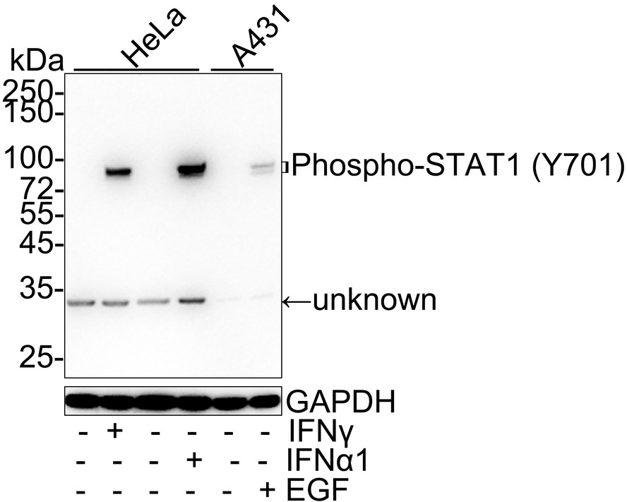
Western blot analysis of Phospho-STAT1 (Y701) on different lysates with Rabbit anti-Phospho-STAT1 (Y701) antibody at 1/1,000 dilution. Lane 1: HeLa cell lysate, Lane 2: HeLa treated with 100ng/mL IFNγ for 30 minutes cell lysate, Lane 3: HeLa cell lysate, Lane 4: HeLa treated with 50ng/mL IFNα1 for 30 minutes cell lysate, Lane 5: A431 cell lysate, Lane 6: A431 treated with 100ng/mL EGF for 30 minutes cell lysate, Lysates/proteins at 20 µg/Lane. Exposure time: 3 minutes; 4-20% SDS-PAGE gel. Proteins were transferred to a PVDF membrane and blocked with 5% NFDM/TBST for 1 hour at room temperature. The primary antibody at 1/1,000 dilution was used in 5% NFDM/TBST at 4℃ overnight. Goat Anti-Rabbit IgG - HRP Secondary Antibody at 1/50,000 dilution was used for 1 hour at room temperature.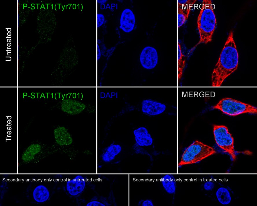
ICC/IF
Immunocytochemistry analysis of HeLa cells treated with 100ng/mL IFNα1 for 5 minutes labeling Phospho-STAT1 (Y701) with Rabbit anti-Phospho-STAT1 (Y701) antibody at 1/100 dilution. Cells were fixed in 4% paraformaldehyde for 20 minutes at room temperature, permeabilized with 0.1% Triton X-100 in PBS for 5 minutes at room temperature, then blocked with 1% BSA in 10% negative goat serum for 1 hour at room temperature. Cells were then incubated with Rabbit anti-Phospho-STAT1 (Y701) antibody at 1/100 dilution in 1% BSA in PBST overnight at 4 ℃. Goat Anti-Rabbit IgG H&L (488) was used as the secondary antibody at 1/1,000 dilution. PBS instead of the primary antibody was used as the secondary antibody only control. Nuclear DNA was labelled in blue with DAPI. Beta tubulin (red) was stained at 1/100 dilution overnight at +4℃. Goat Anti-Mouse IgG H&L (594) was used as the secondary antibody at 1/1,000 dilution.| Application Notes | WB:1:1000 ICC/IF:1:100 |
|---|
| Form | Liquid |
|---|---|
| Storage Instructions | Store at +4℃ after thawing. Aliquot store at -20℃. Avoid repeated freeze / thaw cycles. |
| Storage Buffer | PBS (pH7.4), 0.1% BSA, 40% Glycerol. Preservative: 0.05% Sodium Azide. |
抱歉,暂无相关文献







