| 存储条件 |
|---|
| Product Name | HIF-1 alpha Recombinant Rabbit Monoclonal Antibody |
|---|---|
| Antibody Type | Primary Antibodies |
| Immunogen | Synthetic peptide within human HIF-1 alpha aa 441-490. |
| Clonality | monoclonal |
|---|---|
| Isotype | IgG |
| Host Species | Rabbit |
| Tested Applications | ICC/IFIHCWB |
| WB:1:5000 IHC:1:1000 ICC/IF:1:100 | |
| Species Reactivity | HumanMouse |
| Concentration | 1mg/ml |
| Purification | Protein A |
| Gene Symbol | HIF1A |
|---|---|
| Gene Synonyms | HIF1 MOP1 PASD8 HIF-1A bHLHe78 HIF-1alpha HIF1-ALPHA HIF-1-alpha |
| Gene Full Name | hypoxia inducible factor 1 subunit alpha |
| Gene Summary | This gene encodes the alpha subunit of transcription factor hypoxia-inducible factor-1 (HIF-1), which is a heterodimer composed of an alpha and a beta subunit. HIF-1 functions as a master regulator of cellular and systemic homeostatic response to hypoxia by activating transcription of many genes, including those involved in energy metabolism, angiogenesis, apoptosis, and other genes whose protein products increase oxygen delivery or facilitate metabolic adaptation to hypoxia. HIF-1 thus plays an essential role in embryonic vascularization, tumor angiogenesis and pathophysiology of ischemic disease. Alternatively spliced transcript variants encoding different isoforms have been identified for this gene. [provided by RefSeq, Jul 2011] |
| Alternative Names | ARNT interacting protein antibody ARNT-interacting protein antibody Basic helix loop helix PAS protein MOP1 antibody Basic-helix-loop-helix-PAS protein MOP1 antibody bHLHe78 antibody Class E basic helix-loop-helix protein 78 antibody HIF 1A antibody HIF 1alpha antibody HIF-1-alpha antibody HIF-1alpha antibody ARNT interacting protein antibody ARNT-interacting protein antibody Basic helix loop helix PAS protein MOP1 antibody Basic-helix-loop-helix-PAS protein MOP1 antibody bHLHe78 antibody Class E basic helix-loop-helix protein 78 antibody HIF 1A antibody HIF 1alpha antibody HIF-1-alpha antibody HIF-1alpha antibody HIF-alpha antibody HIF1 A antibody HIF1 Alpha antibody HIF1 antibody HIF1-alpha antibody HIF1A antibody HIF1A_HUMAN antibody hifla antibody Hypoxia inducible factor 1 alpha antibody Hypoxia inducible factor 1 alpha isoform I.3 antibody Hypoxia inducible factor 1 alpha subunit antibody Hypoxia inducible factor 1 alpha subunit basic helix loop helix transcription factor antibody Hypoxia inducible factor 1 alpha subunit (basic helix loop helix transcription factor) antibody Hypoxia inducible factor1alpha antibody Hypoxia-inducible factor 1-alpha antibody Member of PAS protein 1 antibody Member of PAS superfamily 1 antibody Member of the PAS Superfamily 1 antibody MOP 1 antibody MOP1 antibody PAS domain-containing protein 8 antibody PASD 8 antibody PASD8 antibody |
| Molecular Weight(MW) | 93kDa(Observed band size: 120kDa) |
| Cellular Localization | Cytoplasm, Nucleus, Nucleus speckle. |
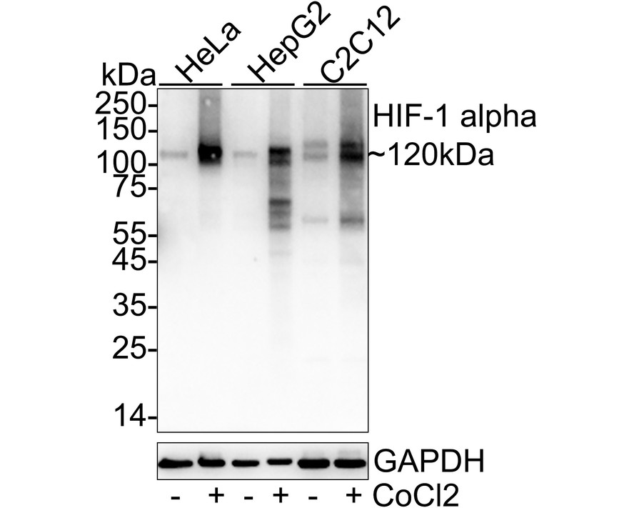
WB
Western blot analysis of HIF-1 alpha on different lysates with Rabbit anti-HIF-1 alpha antibody at 1/5,000 dilution. Lane 1: HeLa cell lysate, Lane 2: HeLa treated with 0.5mM CoCl2 for 6 hours cell lysate, Lane 3: HepG2 cell lysate, Lane 4: HepG2 treated with 100μM CoCl2 for 4 hours cell lysate, Lane 5: C2C12 cell lysate, Lane 6: C2C12 treated with 100μM CoCl2 for 4 hours cell lysate, Lysates/proteins at 30 µg/Lane. Exposure time: 20 seconds; 4-20% SDS-PAGE gel. Proteins were transferred to a PVDF membrane and blocked with 5% NFDM/TBST for 1 hour at room temperature. The primary antibody at 1/5,000 dilution was used in 5% NFDM/TBST at 4℃ overnight. Goat Anti-Rabbit IgG - HRP Secondary Antibody at 1/50,000 dilution was used for 1 hour at room temperature.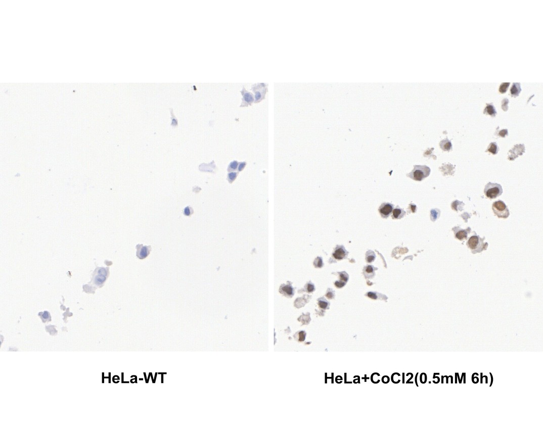
IHC
Immunohistochemical analysis of paraffin-embedded HeLa cells treated with 0.5mM CoCl2 for 6 hours with Rabbit anti-HIF-1 alpha antibody at 1/1,000 dilution. The section was pre-treated using heat mediated antigen retrieval with sodium citrate buffer (pH 6.0) for 2 minutes. The tissues were blocked in 1% BSA for 20 minutes at room temperature, washed with ddH2O and PBS, and then probed with the primary antibody at 1/1,000 dilution for 1 hour at room temperature. The detection was performed using an HRP conjugated compact polymer system. DAB was used as the chromogen. Tissues were counterstained with hematoxylin and mounted with DPX.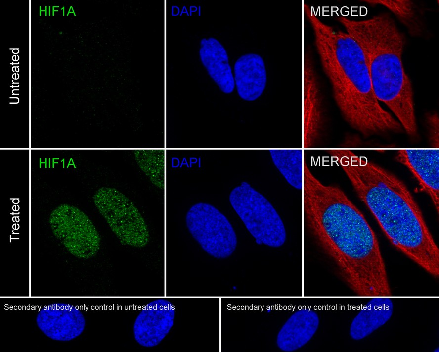
ICC/IF
Immunocytochemistry analysis of HeLa cells treated with 0.5mM CoCl2 for 6 hours labeling HIF-1 alpha with Rabbit anti-HIF-1 alpha antibody at 1/100 dilution. Cells were fixed in 4% paraformaldehyde for 20 minutes at room temperature, permeabilized with 0.1% Triton X-100 in PBS for 5 minutes at room temperature, then blocked with 1% BSA in 10% negative goat serum for 1 hour at room temperature. Cells were then incubated with Rabbit anti-HIF-1 alpha antibody at 1/100 dilution in 1% BSA in PBST overnight at 4 ℃. Goat Anti-Rabbit IgG H&L (iFluor™ 488) was used as the secondary antibody at 1/1,000 dilution. PBS instead of the primary antibody was used as the secondary antibody only control. Nuclear DNA was labelled in blue with DAPI. Beta tubulin (red) was stained at 1/100 dilution overnight at +4℃. Goat Anti-Mouse IgG H&L (iFluor™ 594) was used as the secondary antibody at 1/1,000 dilution.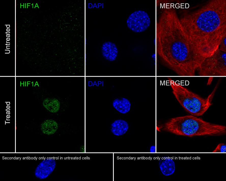
ICC/IF
Immunocytochemistry analysis of C2C12 cells treated with 200μM CoCl2 for 4 hours labeling HIF-1 alpha with Rabbit anti-HIF-1 alpha antibody at 1/100 dilution. Cells were fixed in 4% paraformaldehyde for 20 minutes at room temperature, permeabilized with 0.1% Triton X-100 in PBS for 5 minutes at room temperature, then blocked with 1% BSA in 10% negative goat serum for 1 hour at room temperature. Cells were then incubated with Rabbit anti-HIF-1 alpha antibody at 1/100 dilution in 1% BSA in PBST overnight at 4 ℃. Goat Anti-Rabbit IgG H&L (iFluor™ 488) was used as the secondary antibody at 1/1,000 dilution. PBS instead of the primary antibody was used as the secondary antibody only control. Nuclear DNA was labelled in blue with DAPI. Beta tubulin (red) was stained at 1/100 dilution overnight at +4℃. Goat Anti-Mouse IgG H&L (iFluor™ 594) was used as the secondary antibody at 1/1,000 dilution.
WB
(E) & (F) Western blotting studies of HIF-1α level in lymph nodes and spleen from WT, microRNA 182 OE and microRNA 182 KD mice.| Application Notes | WB:1:5000 IHC:1:1000 ICC/IF:1:100 |
|---|
| Form | Liquid |
|---|---|
| Storage Instructions | Store at +4℃ after thawing. Aliquot store at -20℃. Avoid repeated freeze / thaw cycles. |
| Storage Buffer | PBS (pH7.4), 0.1% BSA, 40% Glycerol. Preservative: 0.05% Sodium Azide. |
抱歉,暂无相关文献







