| 存储条件 |
|---|
| Product Name | S100A9 Rabbit mAb |
|---|---|
| Antibody Type | Primary Antibodies |
| Immunogen | A sRecombinant fusion protein containing a sequence corresponding to amino acids 1-113 of mouse S100a9 (NP_033140.1). |
| Clonality | monoclonal |
|---|---|
| Isotype | IgG |
| Host Species | Rabbit |
| Tested Applications | IF-PIHCWB |
| WB:1:1500-1:12000 IHC:1:200-1:800 IF-P:1:200-1:800 | |
| Species Reactivity | HumanMouseRat |
| Concentration | 1mg/ml |
| Purification | Affinity purified |
| Gene Symbol | S100A9 |
|---|---|
| Gene Synonyms | MIF NIF P14 CAGB CFAG CGLB L1AG LIAG MRP14 60B8AG MAC387 S100-A9 |
| Gene Full Name | S100 calcium binding protein A9 |
| Gene Summary | The protein encoded by this gene is a member of the S100 family of proteins containing 2 EF-hand calcium-binding motifs. S100 proteins are localized in the cytoplasm and/or nucleus of a wide range of cells, and involved in the regulation of a number of cellular processes such as cell cycle progression and differentiation. S100 genes include at least 13 members which are located as a cluster on chromosome 1q21. This protein may function in the inhibition of casein kinase and altered expression of this protein is associated with the disease cystic fibrosis. This antimicrobial protein exhibits antifungal and antibacterial activity. [provided by RefSeq, Nov 2014] |
| Alternative Names | MIF NIF P14 CAGB CFAG CGLB L1AG LIAG MRP14 60B8AG MAC387 S100-A9 |
| Molecular Weight(MW) | 14kDa |
| Cellular Localization | Secreted, Cytoplasm, Cell membrane , Cell membrane, Peripheral membrane protein, cytoskeleton, cell junction, cytoplasm, cytosol, extracellular exosome, extracellular region, extracellular space, nucleoplasm, nucleus, plasma membrane. |
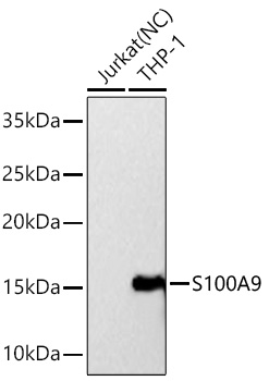
WB
Western blot analysis of various lysates using S100A9 Rabbit mAb at 1:3000 dilution incubated overnight at 4℃. Secondary antibody: HRP-conjugated Goat anti-Rabbit IgG (H+L) at 1:10000 dilution. Lysates/proteins: 25μg per lane. Blocking buffer: 3% nonfat dry milk in TBST. Detection: ECL Basic Kit. Negative control (NC): Jurkat Exposure time: 90s.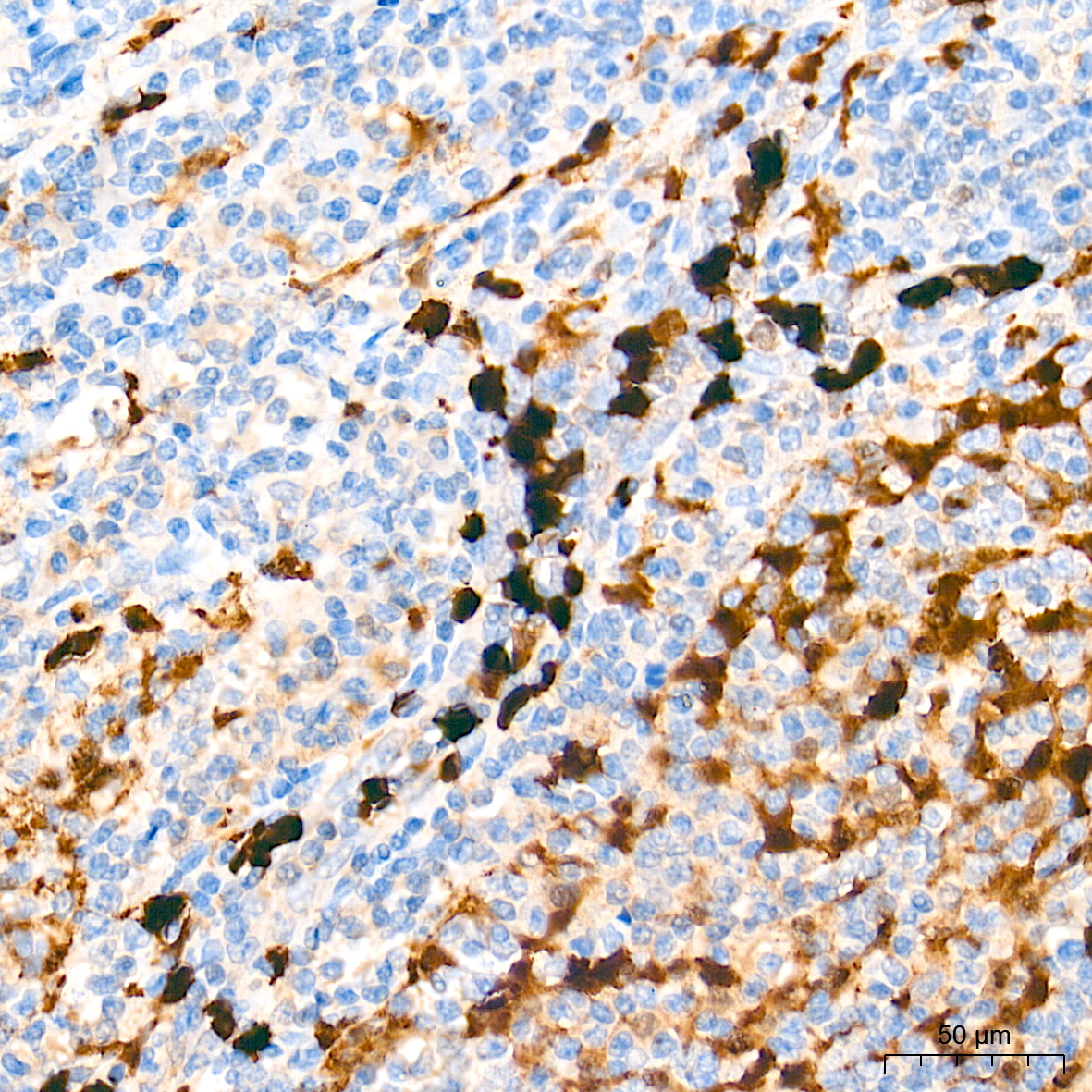
IHC
Immunohistochemistry analysis of paraffin embedded Human tonsil tissue using S100A9 Rabbit mAb at a dilution of 1:500 (40x lens). High pressure antigen retrieval performed with 0.01M Tris-EDTA Buffer (pH 9.0) prior to IHC staining.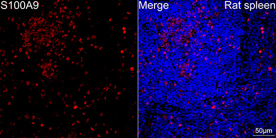
IF-P
Confocal imaging of paraffin-embedded Rat spleen tissue using S100A9 Rabbit mAb (dilution 1:200) followed by a further incubation with Cy3 Goat Anti-Rabbit IgG (H+L)(dilution 1:500) (Red). DAPI was used for nuclear staining (Blue). High pressure antigen retrieval performed with 0.01M Citrate Buffer (pH 6.0) prior to IF staining. Objective: 40x.| Application Notes | WB:1:1500-1:12000 IHC:1:200-1:800 IF-P:1:200-1:800 |
|---|
| Form | Liquid |
|---|---|
| Storage Instructions | Store at -20℃. Avoid freeze / thaw cycles. |
| Storage Buffer | Buffer: PBS with 0.05% proclin300, 0.05% BSA, 50% glycerol, pH7.3. |
抱歉,暂无相关文献







