| 存储条件 |
|---|
| Product Name | Phospho-EIF2S1 (S51) Recombinant Rabbit Monoclonal Antibody |
|---|---|
| Antibody Type | Primary Antibodies |
| Immunogen | Synthetic phospho-peptide corresponding to residues surrounding Ser51 of Human eIF-2a. |
| Modification | p-S51 |
| Clonality | monoclonal |
|---|---|
| Isotype | IgG |
| Host Species | Rabbit |
| Tested Applications | FCIHCWB |
| WB:1:2000 IHC:1:50-1:500 FC:1:50-1:100 | |
| Species Reactivity | HumanMouseRat |
| Concentration | 1mg/ml |
| Purification | Protein A |
| Gene Symbol | EIF2S1 |
|---|---|
| Gene Synonyms | EIF2 EIF-2 EIF2A EIF-2A EIF-2alpha |
| Gene Full Name | eukaryotic translation initiation factor 2 subunit alpha |
| Gene Summary | The translation initiation factor EIF2 catalyzes the first regulated step of protein synthesis initiation, promoting the binding of the initiator tRNA to 40S ribosomal subunits. Binding occurs as a ternary complex of methionyl-tRNA, EIF2, and GTP. EIF2 is composed of 3 nonidentical subunits, the 36-kD EIF2-alpha subunit (EIF2S1), the 38-kD EIF2-beta subunit (EIF2S2; MIM 603908), and the 52-kD EIF2-gamma subunit (EIF2S3; MIM 300161). The rate of formation of the ternary complex is modulated by the phosphorylation state of EIF2-alpha (Ernst et al., 1987 [PubMed 2948954]).[supplied by OMIM, Feb 2010]. |
| Alternative Names | EIF 2 alpha antibody EIF 2 antibody EIF 2A antibody EIF 2alpha antibody eIF-2-alpha antibody eIF-2A antibody EIF-2alpha antibody EIF2 alpha antibody EIF2 antibody EIF2A antibody EIF 2 alpha antibody EIF 2 antibody EIF 2A antibody EIF 2alpha antibody eIF-2-alpha antibody eIF-2A antibody EIF-2alpha antibody EIF2 alpha antibody EIF2 antibody EIF2A antibody EIF2S1 antibody Eukaryotic translation initiation factor 2 subunit 1 alpha 35kDa antibody Eukaryotic translation initiation factor 2 subunit 1 alpha antibody Eukaryotic translation initiation factor 2 subunit 1 antibody Eukaryotic translation initiation factor 2 subunit alpha antibody IF2A_HUMAN antibody |
| Molecular Weight(MW) | 36kDa |
| Function | Phosphorylation of the eukaryotic initiation factor 2 (eIF2) α subunit is a well-documented mechanism to downregulate protein synthesis under a variety of stress conditions. Eukaryotic initiation factor 2 binds GTP and Met-tRNAi and transfers Met-tRNA to the 40S subunit to form the 43S preinitiation complex. eIF2 promotes a new round of translation initiation by exchanging GDP for GTP, a reaction catalyzed by eIF2B. Kinases that are activated by viral infection (PKR), endoplasmic reticulum stress (PERK/PEK), amino acid deprivation (GCN2), or heme deficiency (HRI) can phosphorylate the α subunit of eIF2. This phosphorylation stabilizes the eIF2-GDP-eIF2B complex and inhibits the turnover of eIF2B. Induction of PKR by IFN-γ and TNF-α induces potent phosphorylation of eIF2α at Ser51. |
| Cellular Localization | Cytoplasm, Stress granule. |
WB
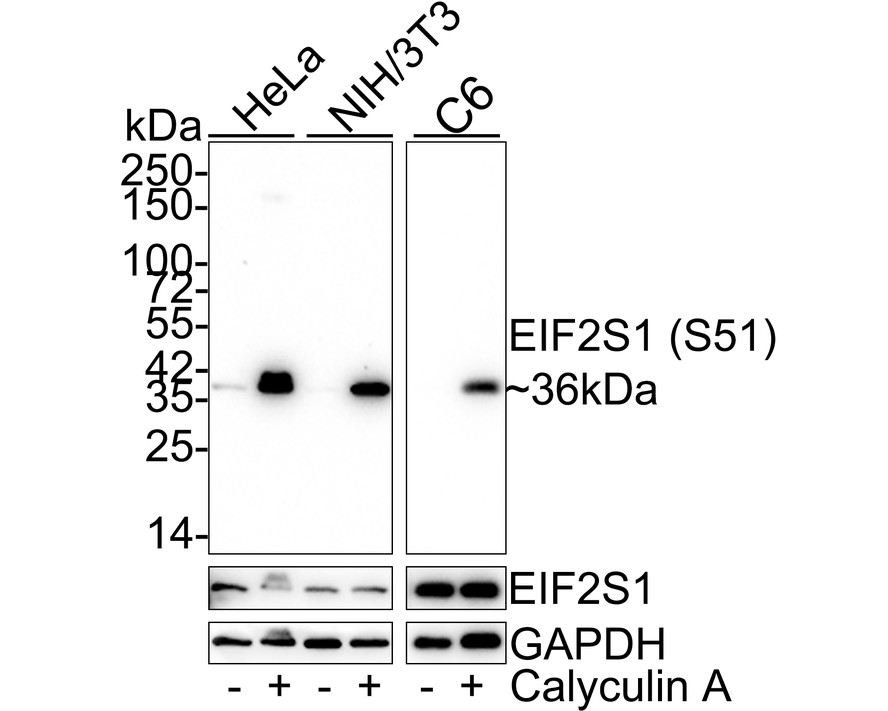
Western blot analysis of Phospho-EIF2S1 (S51) on different lysates with Rabbit anti-Phospho-EIF2S1 (S51) antibody at 1/2,000 dilution. Lane 1: HeLa whole cell lysate (15 µg/Lane), Lane 2: HeLa treated with 50nM Calyculin A for 3 hours whole cell lysate (15 µg/Lane), Lane 3: NIH/3T3 whole cell lysate (15 µg/Lane), Lane 4: NIH/3T3 treated with 100nM Calyculin A for 30 minutes whole cell lysate (15 µg/Lane), Lane 5: C6 whole cell lysate (20 µg/Lane), Lane 6: C6 treated with 100nM Calyculin A for 30 minutes whole cell lysate (20 µg/Lane), Exposure time: Lane 1-4: 2 minutes; Lane 5-6: 23 seconds; 4-20% SDS-PAGE gel. Proteins were transferred to a PVDF membrane and blocked with 5% NFDM/TBST for 1 hour at room temperature. The primary antibody at 1/2,000 dilution was used in 5% NFDM/TBST at 4℃ overnight. Goat Anti-Rabbit IgG - HRP Secondary Antibody at 1:50,000 dilution was used for 1 hour at room temperature.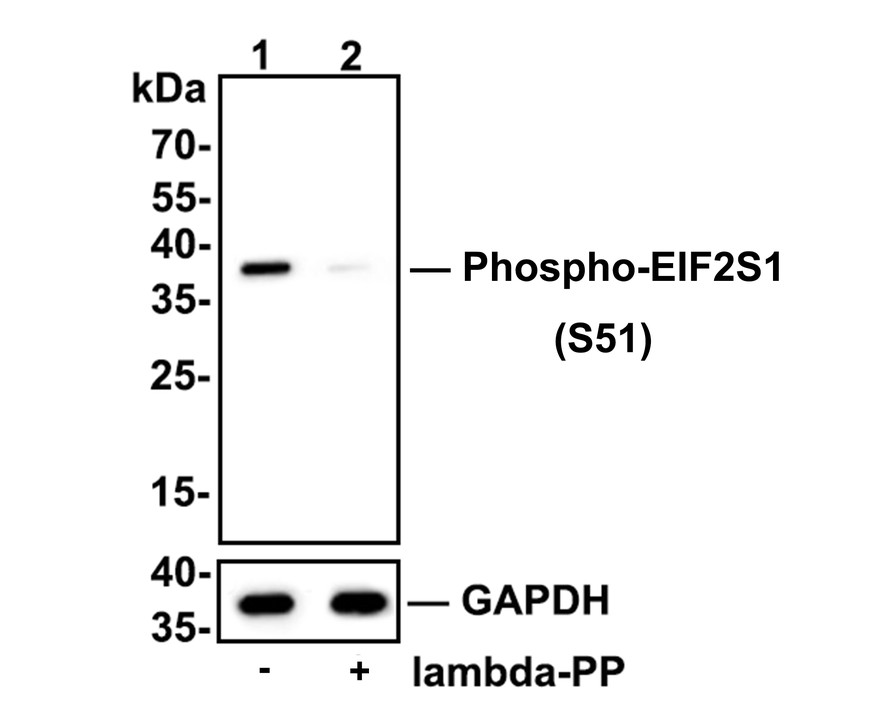
WB
Western blot analysis of Phospho-EIF2S1 (S51) on mouse spleen tissue lysates. Lane 1: mouse spleen tissue, whole tissue lysate, 20ug/lane. Lane 2: mouse spleen tissue treated with 2.8ug/ul lambda-PP for 30 minutes, whole tissue lysates, 20ug/lane. All lanes : Anti-Phospho-EIF2S1 (S51) antibody at 1/500 dilution. Anti-GAPDH antibody at 1/10,000 dilution. Goat Anti-Rabbit IgG H&L (HRP) at 1/200,000 dilution. Blocking and diluting buffer: 5% BSA. Exposure time: 3 minutes 43 seconds.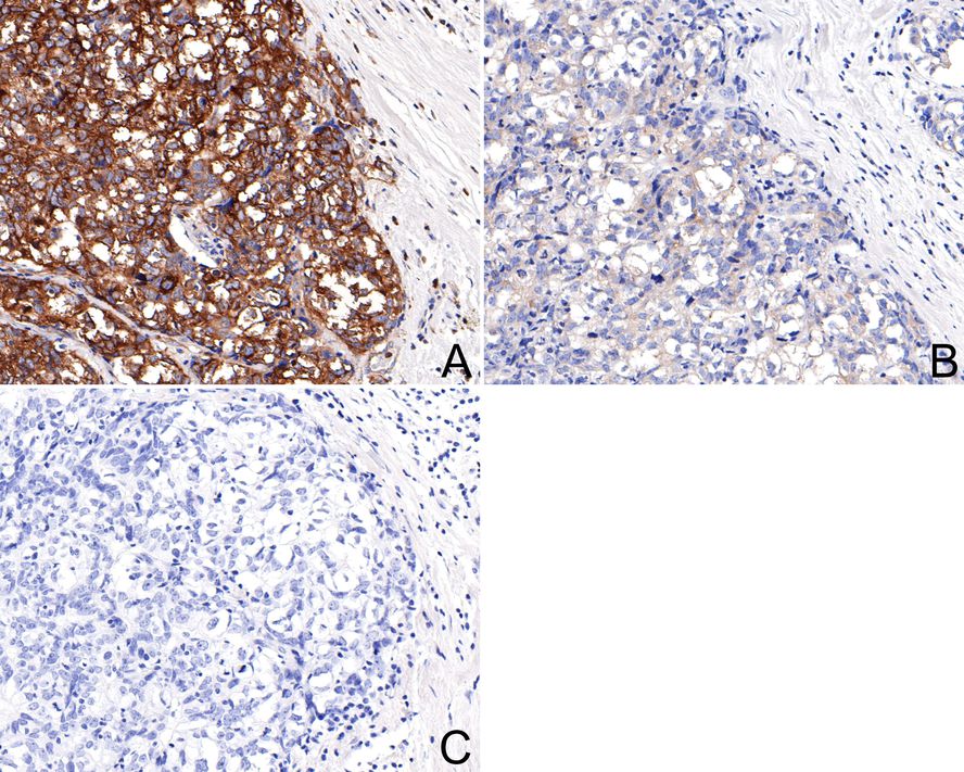
IHC
Immunohistochemical analysis of paraffin-embedded human breast carcinoma tissue with Rabbit anti-Phospho-EIF2S1 (S51) antibody at 1/200 dilution. A: Untreated human breast carcinoma tissue, B: λ-PPase treated human breast carcinoma tissue, C: Negative control. The section was pre-treated using heat mediated antigen retrieval with Tris-EDTA buffer (pH 9.0) for 20 minutes. The tissues were blocked in 1% BSA for 20 minutes at room temperature, washed with ddH2O and PBS, and then probed with the primary antibody at 1/200 dilution for 1 hour at room temperature. The detection was performed using an HRP conjugated compact polymer system. DAB was used as the chromogen. Tissues were counterstained with hematoxylin and mounted with DPX.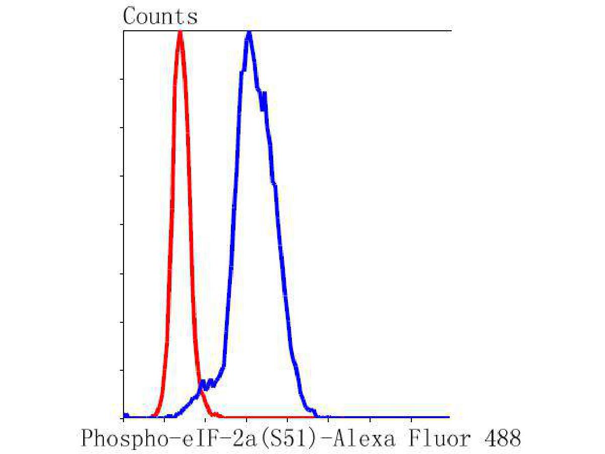
FC
Flow cytometric analysis of Phospho-EIF2S1 (S51) was done on Hela cells. The cells were fixed, permeabilized and stained with the primary antibody (1/50) (blue). After incubation of the primary antibody at room temperature for an hour, the cells were stained with a Alexa Fluor 488-conjugated Goat anti-Rabbit IgG Secondary antibody at 1/1000 dilution for 30 minutes.Unlabelled sample was used as a control (cells without incubation with primary antibody; red).| Application Notes | WB:1:2000 IHC:1:50-1:500 FC:1:50-1:100 |
|---|
| Form | Liquid |
|---|---|
| Storage Instructions | Store at +4℃ after thawing. Aliquot store at -20℃ or -80℃. Avoid repeated freeze / thaw cycles. |
| Storage Buffer | 1*TBS (pH7.4), 0.05% BSA, 40% Glycerol. Preservative: 0.05% Sodium Azide. |
抱歉,暂无相关文献







