| 存储条件 |
|---|
| Product Name | EEA1 Recombinant Rabbit Monoclonal Antibody |
|---|---|
| Antibody Type | Primary Antibodies |
| Immunogen | Recombinant protein within Human EEA1 aa 1-100 / 1,411. |
| Clonality | monoclonal |
|---|---|
| Isotype | IgG |
| Host Species | Rabbit |
| Tested Applications | FCICC/IFWB |
| WB:1:1000 ICC/IF:1:100 FC:1:1000 | |
| Species Reactivity | HumanMonkeyMouseRat |
| Concentration | 1mg/ml |
| Purification | Protein A |
| Gene Symbol | EEA1 |
|---|---|
| Gene Synonyms | MST105 ZFYVE2 MSTP105 |
| Gene Full Name | early endosome antigen 1 |
| Gene Summary | Enables 1-phosphatidylinositol binding activity; GTP-dependent protein binding activity; and protein homodimerization activity. Involved in endocytosis and vesicle fusion. Located in cytosol and early endosome membrane. [provided by Alliance of Genome Resources, Feb 2025] |
| Alternative Names | Early endosome antigen 1 antibody Early endosome antigen 1 162kD antibody Early endosome associated protein antibody EEA 1 antibody EEA1 antibody EEA1_HUMAN antibody Endosome associated protein p162 antibody Endosome-associated protein p162 antibody MST105 antibody MSTP105 antibody Early endosome antigen 1 antibody Early endosome antigen 1 162kD antibody Early endosome associated protein antibody EEA 1 antibody EEA1 antibody EEA1_HUMAN antibody Endosome associated protein p162 antibody Endosome-associated protein p162 antibody MST105 antibody MSTP105 antibody ZFYVE2 antibody Zinc finger FYVE domain containing protein 2 antibody Zinc finger FYVE domain-containing protein 2 antibody |
| Molecular Weight(MW) | 162kDa |
| Cellular Localization | Cytoplasm, Early endosome membrane. |
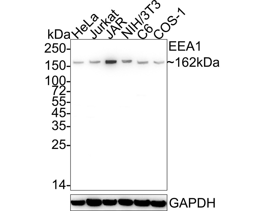
WB
Western blot analysis of EEA1 on different lysates with Rabbit anti-EEA1 antibody at 1/1,000 dilution. Lane 1: HeLa cell lysate, Lane 2: Jurkat cell lysate, Lane 3: JAR cell lysate, Lane 4: NIH/3T3 cell lysate, Lane 5: C6 cell lysate, Lane 6: COS-1 cell lysate, Lysates/proteins at 20 µg/Lane. Exposure time: 12 seconds; 4-20% SDS-PAGE gel. Proteins were transferred to a PVDF membrane and blocked with 5% NFDM/TBST for 1 hour at room temperature. The primary antibody at 1/1,000 dilution was used in 5% NFDM/TBST at 4℃ overnight. Goat Anti-Rabbit IgG - HRP Secondary Antibody at 1/50,000 dilution was used for 1 hour at room temperature.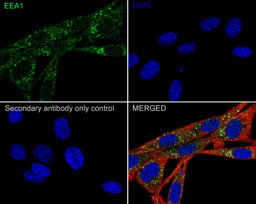
ICC/IF
Immunocytochemistry analysis of NIH/3T3 cells labeling EEA1 with Rabbit anti-EEA1 antibody at 1/100 dilution. Cells were fixed in 4% paraformaldehyde for 20 minutes at room temperature, permeabilized with 0.1% Triton X-100 in PBS for 5 minutes at room temperature, then blocked with 1% BSA in 10% negative goat serum for 1 hour at room temperature. Cells were then incubated with Rabbit anti-EEA1 antibody at 1/100 dilution in 1% BSA in PBST overnight at 4 ℃. Goat Anti-Rabbit IgG H&L (iFluor™ 48) was used as the secondary antibody at 1/1,000 dilution. PBS instead of the primary antibody was used as the secondary antibody only control. Nuclear DNA was labelled in blue with DAPI. Beta tubulin (red) was stained at 1/100 dilution overnight at +4℃. Goat Anti-Mouse IgG H&L (iFluor™ 594) was used as the secondary antibody at 1/1,000 dilution.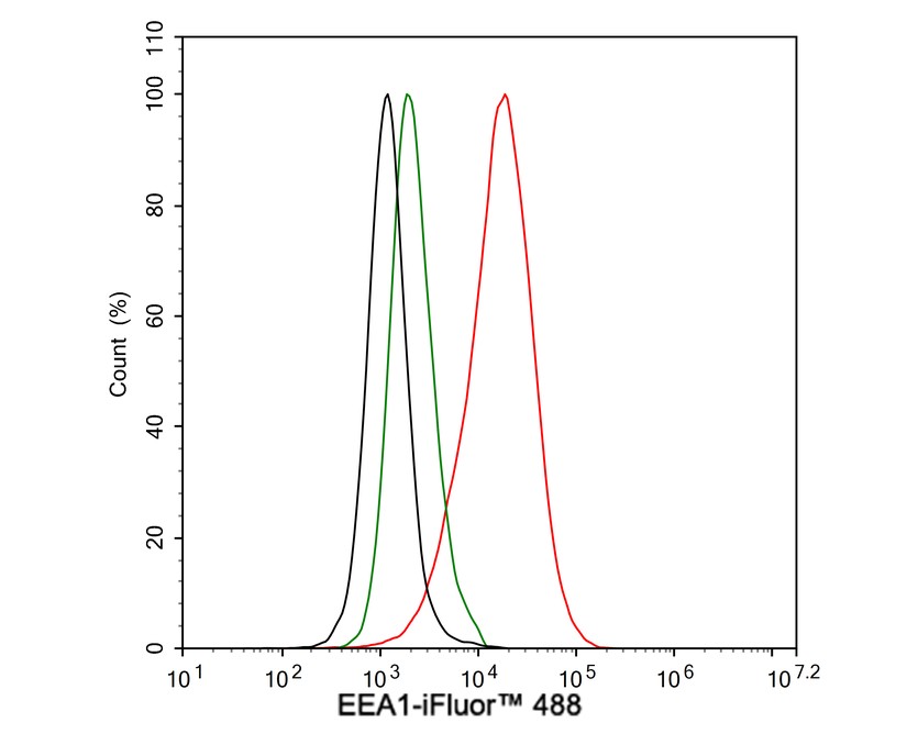
FC
Flow cytometric analysis of JAR cells labeling EEA1. Cells were fixed and permeabilized. Then stained with the primary antibody (1μg/mL) (red) compared with Rabbit IgG Isotype Control (green). After incubation of the primary antibody at +4℃ for an hour, the cells were stained with a iFluor™ 488 conjugate-Goat anti-Rabbit IgG Secondary antibody at 1/1,000 dilution for 30 minutes at +4℃. Unlabelled sample was used as a control (cells without incubation with primary antibody; black).| Application Notes | WB:1:1000 ICC/IF:1:100 FC:1:1000 |
|---|
| Form | Liquid |
|---|---|
| Storage Instructions | Store at +4℃ after thawing. Aliquot store at -20℃ or -80℃. Avoid repeated freeze / thaw cycles. |
| Storage Buffer | 1*TBS (pH7.4), 0.05% BSA, 40% Glycerol. Preservative: 0.05% Sodium Azide. |
抱歉,暂无相关文献







