| 存储条件 |
|---|
| Product Name | Phospholipase C gamma 1 Recombinant Rabbit Monoclonal Antibody |
|---|---|
| Antibody Type | Primary Antibodies |
| Immunogen | Recombinant protein within Human Phospholipase C gamma 1 aa 690-852 / 1,290. |
| Clonality | Monoclonal |
|---|---|
| Isotype | IgG |
| Host Species | Rabbit |
| Tested Applications | ICC/IFIHCWB |
| WB:1:1000 IHC:1:200 ICC/IF:1:50-1:200 | |
| Species Reactivity | HumanMouseRat |
| Concentration | 1mg/ml |
| Purification | Protein A |
| Gene Symbol | PLCG1 |
|---|---|
| Gene Synonyms | IDAA PLC1 NCKAP3 PLC-II PLC148 PLCgamma1 |
| Gene Full Name | phospholipase C gamma 1 |
| Gene Summary | The protein encoded by this gene catalyzes the formation of inositol 1,4,5-trisphosphate and diacylglycerol from phosphatidylinositol 4,5-bisphosphate. This reaction uses calcium as a cofactor and plays an important role in the intracellular transduction of receptor-mediated tyrosine kinase activators. For example, when activated by SRC, the encoded protein causes the Ras guanine nucleotide exchange factor RasGRP1 to translocate to the Golgi, where it activates Ras. Also, this protein has been shown to be a major substrate for heparin-binding growth factor 1 (acidic fibroblast growth factor)-activated tyrosine kinase. Two transcript variants encoding different isoforms have been found for this gene. [provided by RefSeq, Jul 2008] |
| Alternative Names | 1 phosphatidyl D myo inositol 4 5 bisphosphate antibody 1 phosphatidylinositol 4 5 bisphosphate phosphodiesterase gamma 1 antibody 1-phosphatidylinositol-4 5-bisphosphate phosphodiesterase gamma-1 antibody Inositoltrisphosphohydrolase antibody Monophosphatidylinositol phosphodiesterase antibody NCKAP3 antibody Phosphatidylinositol phospholipase C antibody Phosphoinositidase C antibody Phosphoinositide phospholipase C antibody Phosphoinositide phospholipase C-gamma-1 antibody 1 phosphatidyl D myo inositol 4 5 bisphosphate antibody 1 phosphatidylinositol 4 5 bisphosphate phosphodiesterase gamma 1 antibody 1-phosphatidylinositol-4 5-bisphosphate phosphodiesterase gamma-1 antibody Inositoltrisphosphohydrolase antibody Monophosphatidylinositol phosphodiesterase antibody NCKAP3 antibody Phosphatidylinositol phospholipase C antibody Phosphoinositidase C antibody Phosphoinositide phospholipase C antibody Phosphoinositide phospholipase C-gamma-1 antibody Phospholipase C 148 antibody Phospholipase C gamma 1 antibody Phospholipase C-gamma-1 antibody Phospholipase C-II antibody PLC gamma 1 antibody PLC II antibody PLC-148 antibody PLC-gamma-1 antibody PLC-II antibody PLC1 antibody PLC148 antibody Plcg1 antibody PLCG1_HUMAN antibody PLCgamma1 antibody |
| Molecular Weight(MW) | 149kDa |
| Cellular Localization | Cell projection. |
WB
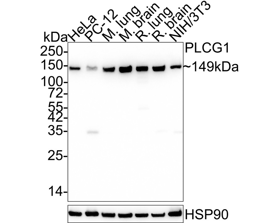
Western blot analysis of Phospholipase C gamma 1 on different lysates with Rabbit anti-Phospholipase C gamma 1 antibody at 1/1,000 dilution. Lane 1: HeLa cell lysate (20 µg/Lane) Lane 2: PC-12 cell lysate (20 µg/Lane) Lane 3: Mouse lung tissue lysate (40 µg/Lane) Lane 4: Mouse brain tissue lysate (40 µg/Lane) Lane 5: Rat lung tissue lysate (40 µg/Lane) Lane 6: Rat brain tissue lysate (40 µg/Lane) Lane 7: NIH/3T3 cell lysate (20 µg/Lane) Exposure time: 50 seconds; 4-20% SDS-PAGE gel. Proteins were transferred to a PVDF membrane and blocked with 5% NFDM/TBST for 1 hour at room temperature. The primary antibody at 1/1,000 dilution was used in 5% NFDM/TBST at 4℃ overnight. Goat Anti-Rabbit IgG - HRP Secondary Antibody at 1/50,000 dilution was used for 1 hour at room temperature.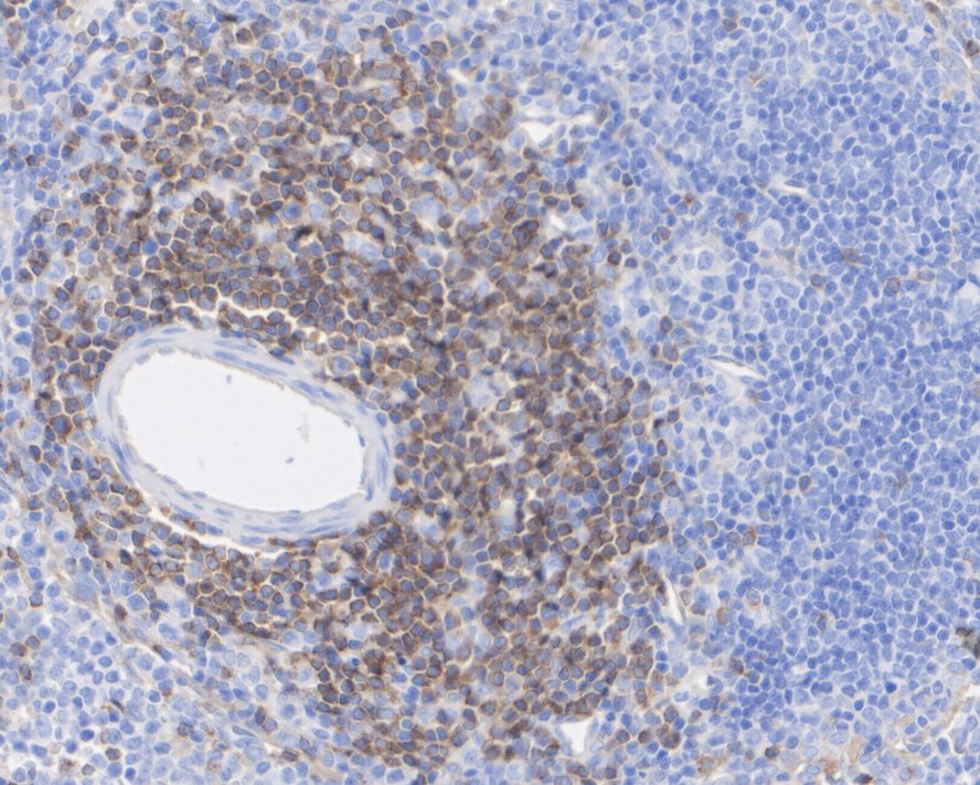
IHC
Immunohistochemical analysis of paraffin-embedded rat spleen tissue with Rabbit anti-Phospholipase C gamma 1 antibody at 1/200 dilution. The section was pre-treated using heat mediated antigen retrieval with Tris-EDTA buffer (pH 9.0) for 20 minutes. The tissues were blocked in 1% BSA for 20 minutes at room temperature, washed with ddH2O and PBS, and then probed with the primary antibody at 1/200 dilution for 1 hour at room temperature. The detection was performed using an HRP conjugated compact polymer system. DAB was used as the chromogen. Tissues were counterstained with hematoxylin and mounted with DPX.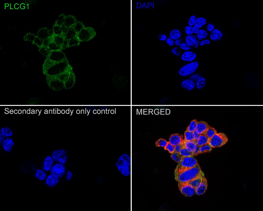
ICC/IF
Immunocytochemistry analysis of PC-12 cells labeling Phospholipase C gamma 1 with Rabbit anti-Phospholipase C gamma 1 antibody at 1/50 dilution. Cells were fixed in 4% paraformaldehyde for 20 minutes at room temperature, permeabilized with 0.1% Triton X-100 in PBS for 5 minutes at room temperature, then blocked with 1% BSA in 10% negative goat serum for 1 hour at room temperature. Cells were then incubated with Rabbit anti-Phospholipase C gamma 1 antibody at 1/50 dilution in 1% BSA in PBST overnight at 4 ℃. Goat Anti-Rabbit IgG H&L (iFluor™ 488) was used as the secondary antibody at 1/1,000 dilution. PBS instead of the primary antibody was used as the secondary antibody only control. Nuclear DNA was labelled in blue with DAPI. Beta tubulin (red) was stained at 1/100 dilution overnight at +4℃. Goat Anti-Mouse IgG H&L (iFluor™ 594) was used as the secondary antibody at 1/1,000 dilution.| Application Notes | WB:1:1000 IHC:1:200 ICC/IF:1:50-1:200 |
|---|
| Form | Liquid |
|---|---|
| Storage Instructions | Store at +4℃ after thawing. Aliquot store at -20℃ or -80℃. Avoid repeated freeze / thaw cycles. |
| Storage Buffer | 1*TBS (pH7.4), 0.05% BSA, 40% Glycerol. Preservative: 0.05% Sodium Azide. |
抱歉,暂无相关文献







