| 存储条件 |
|---|
| Product Name | PCSK9 Rabbit mAb |
|---|---|
| Antibody Type | Primary Antibodies |
| Immunogen | Recombinant fusion protein containing a sequence corresponding to amino acids 31-692 of human PCSK9 (NP_777596.2). |
| Clonality | Monoclonal |
|---|---|
| Isotype | IgG |
| Host Species | Rabbit |
| Tested Applications | ICC/IFIHCWB |
| WB:1:2000-1:12000 IHC:1:200-1:2000 ICC:1:200-1:2000 | |
| Species Reactivity | HumanMouse |
| Concentration | 1mg/ml |
| Purification | Affinity purified |
| Gene Symbol | PCSK9 |
|---|---|
| Gene Synonyms | FH3 PC9 FHCL3 NARC1 LDLCQ1 NARC-1 HCHOLA3 |
| Gene Full Name | proprotein convertase subtilisin/kexin type 9 |
| Gene Summary | This gene encodes a member of the subtilisin-like proprotein convertase family, which includes proteases that process protein and peptide precursors trafficking through regulated or constitutive branches of the secretory pathway. The encoded protein undergoes an autocatalytic processing event with its prosegment in the ER and is constitutively secreted as an inactive protease into the extracellular matrix and trans-Golgi network. It is expressed in liver, intestine and kidney tissues and escorts specific receptors for lysosomal degradation. It plays a role in cholesterol and fatty acid metabolism. Mutations in this gene have been associated with autosomal dominant familial hypercholesterolemia. Alternative splicing results in multiple transcript variants. [provided by RefSeq, Feb 2014] |
| Alternative Names | PCSK9 FH3 HCHOLA3 LDLCQ1 NARC-1 NARC1 PC9 proprotein convertase subtilisin/kexin type 9 |
| Molecular Weight(MW) | 20kDa/74kDa(Observed MW 65kDa,80kDa) |
| Cellular Localization | Cell surface, Cytoplasm, Endoplasmic reticulum, Endosome, Golgi apparatus, Lysosome, Secreted. |
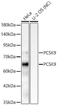
WB
Western blot analysis of various lysates using PCSK9 Rabbit mAb at 1:2000 dilution. Secondary antibody: HRP-conjugated Goat anti-Rabbit IgG (H+L) at 1:10000 dilution. Lysates/proteins: 25 μg per lane. Blocking buffer: 3% nonfat dry milk in TBST. Detection: ECL Basic Kit. Negative control (NC): U-2OS. Exposure time: 10s.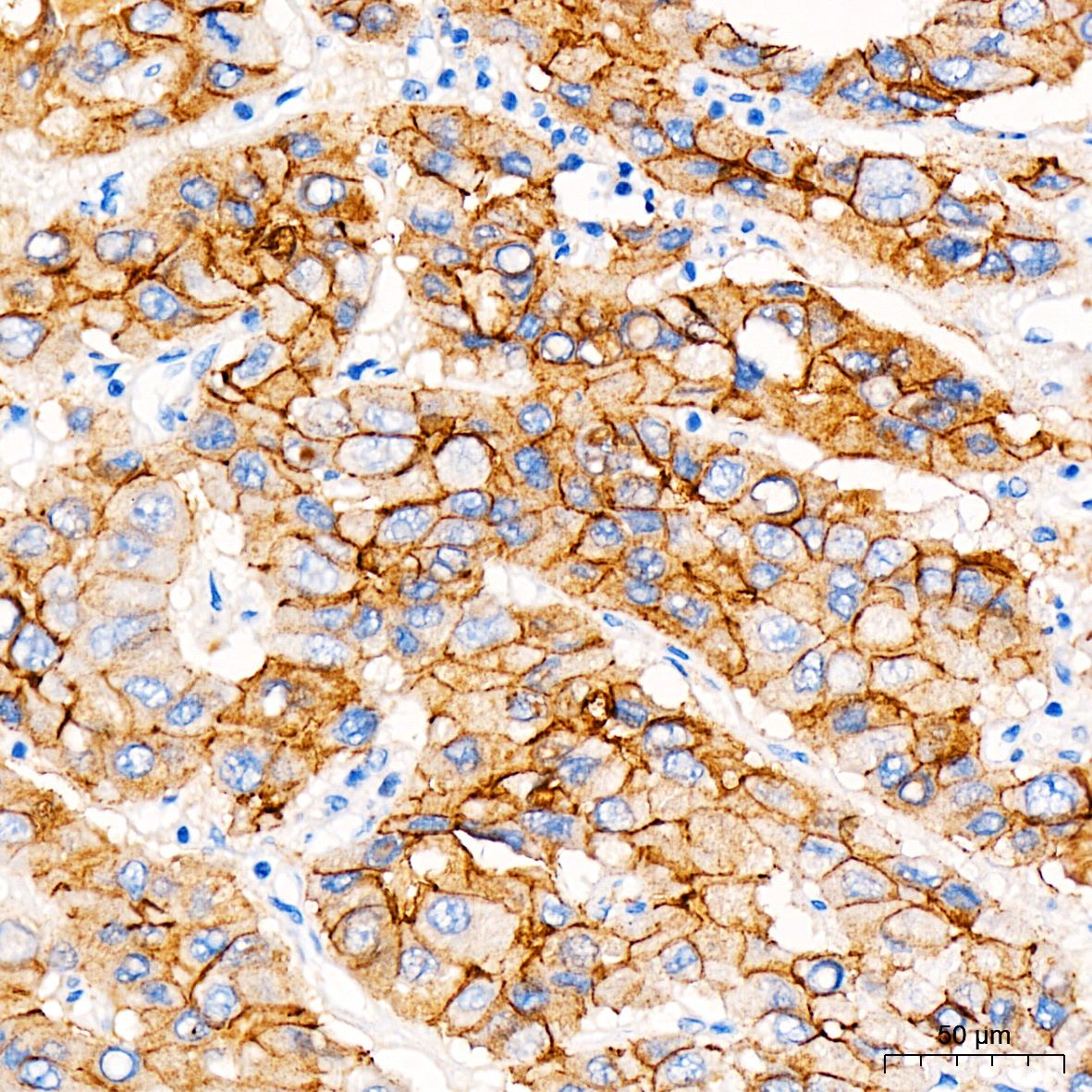
IHC
Immunohistochemistry analysis of paraffin embedded Human liver cancer tissue using PCSK9 Rabbit mAb at a dilution of 1:800 (40x lens). High pressure antigen retrieval was performed with 0.01 M citrate buffer (pH 6.0) prior to IHC staining.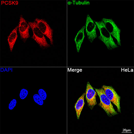
ICC/IF
Confocal imaging of HeLa cells using PCSK9 Rabbit mAb (dilution 1:200) followed by a further incubation with Cy3 Goat Anti-Rabbit IgG (H+L) (dilution 1:500) (Red). The cells were counterstained with α-Tubulin Mouse mAb (dilution 1:400) followed by incubation with Ominimabs® 488-conjugated Goat Anti-Mouse IgG (H+L) Ab (dilution 1:500) (Green). DAPI was used for nuclear staining (Blue). objective: 100x.| Application Notes | WB:1:2000-1:12000 IHC:1:200-1:2000 ICC:1:200-1:2000 |
|---|
| Form | Liquid |
|---|---|
| Storage Instructions | Store at -20℃. Avoid freeze / thaw cycles. |
| Storage Buffer | Buffer: PBS with 0.05% proclin300, 0.05% BSA, 50% glycerol, pH7.3. |
抱歉,暂无相关文献







