| 存储条件 |
|---|
| Product Name | COL1A2 Recombinant Rabbit Monoclonal Antibody |
|---|---|
| Antibody Type | Primary Antibodies |
| Immunogen | Recombinant protein within human COL1A2 aa 1,081-1,366. |
| Clonality | Monoclonal |
|---|---|
| Isotype | IgG |
| Host Species | Rabbit |
| Tested Applications | FCICC/IFWB |
| WB:1:2000 ICC:1:100 FC:1:1000 | |
| Species Reactivity | HumanRat |
| Concentration | 1mg/ml |
| Purification | Protein A |
| Gene Symbol | COL1A2 |
|---|---|
| Gene Synonyms | OI4 EDSCV EDSARTH2 |
| Gene Full Name | collagen type I alpha 2 chain |
| Gene Summary | This gene encodes the pro-alpha2 chain of type I collagen whose triple helix comprises two alpha1 chains and one alpha2 chain. Type I is a fibril-forming collagen found in most connective tissues and is abundant in bone, cornea, dermis and tendon. Mutations in this gene are associated with osteogenesis imperfecta types I-IV, Ehlers-Danlos syndrome type VIIB, recessive Ehlers-Danlos syndrome Classical type, idiopathic osteoporosis, and atypical Marfan syndrome. Symptoms associated with mutations in this gene, however, tend to be less severe than mutations in the gene for the alpha1 chain of type I collagen (COL1A1) reflecting the different role of alpha2 chains in matrix integrity. Three transcripts, resulting from the use of alternate polyadenylation signals, have been identified for this gene. [provided by R. Dalgleish, Feb 2008] |
| Alternative Names | Alpha 2 collagen type I antibody Alpha 2 type I collagen antibody Alpha 2 type I procollagen antibody Alpha 2(I) collagen antibody Alpha 2(I) procollagen antibody Alpha-2 type I collagen antibody CO1A2_HUMAN antibody COL1A2 antibody Collagen alpha 2(I) chain antibody Collagen alpha-2(I) chain antibody Alpha 2 collagen type I antibody Alpha 2 type I collagen antibody Alpha 2 type I procollagen antibody Alpha 2(I) collagen antibody Alpha 2(I) procollagen antibody Alpha-2 type I collagen antibody CO1A2_HUMAN antibody COL1A2 antibody Collagen alpha 2(I) chain antibody Collagen alpha-2(I) chain antibody Collagen I alpha 2 polypeptide antibody Collagen of skin tendon and bone alpha 2 chain antibody Collagen type I alpha 2 antibody OI4 antibody Osteogenesis imperfecta type IV antibody Type I procollagen antibody |
| Molecular Weight(MW) | 129kDa(Observed band size: 150kDa) |
| Cellular Localization | Secreted, extracellular space, extracellular matrix. |
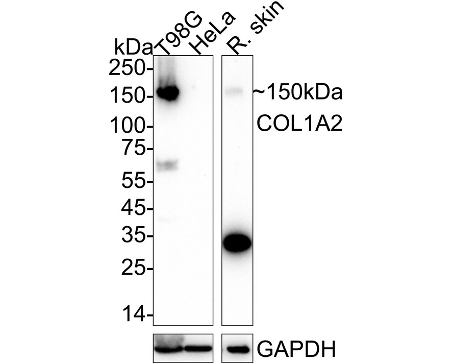
WB
Western blot analysis of COL1A2 on different lysates with Rabbit anti-COL1A2 antibody at 1/2,000 dilution. Lane 1: T98G cell lysate (20 µg/Lane) Lane 2: HeLa cell lysate (negative) (20 µg/Lane) Lane 3: Rat skin tissue lysate (40 µg/Lane) Exposure time: 3 minutes; 4-20% SDS-PAGE gel. Proteins were transferred to a PVDF membrane and blocked with 5% NFDM/TBST for 1 hour at room temperature. The primary antibody at 1/2,000 dilution was used in 5% NFDM/TBST at 4℃ overnight. Goat Anti-Rabbit IgG - HRP Secondary Antibody at 1/50,000 dilution was used for 1 hour at room temperature.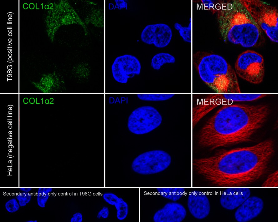
ICC/IF
Immunocytochemistry analysis of T98G (positive) and HeLa (negative) labeling COL1A2 with Rabbit anti-COL1A2 antibody at 1/100 dilution. Cells were fixed in 4% paraformaldehyde for 20 minutes at room temperature, permeabilized with 0.1% Triton X-100 in PBS for 5 minutes at room temperature, then blocked with 1% BSA in 10% negative goat serum for 1 hour at room temperature. Cells were then incubated with Rabbit anti-COL1A2 antibody at 1/100 dilution in 1% BSA in PBST overnight at 4 ℃. Goat Anti-Rabbit IgG H&L (iFluor™ 488) was used as the secondary antibody at 1/1,000 dilution. PBS instead of the primary antibody was used as the secondary antibody only control. Nuclear DNA was labelled in blue with DAPI. Beta tubulin (red) was stained at 1/100 dilution overnight at +4℃. Goat Anti-Mouse IgG H&L (iFluor™ 594) was used as the secondary antibody at 1/1,000 dilution.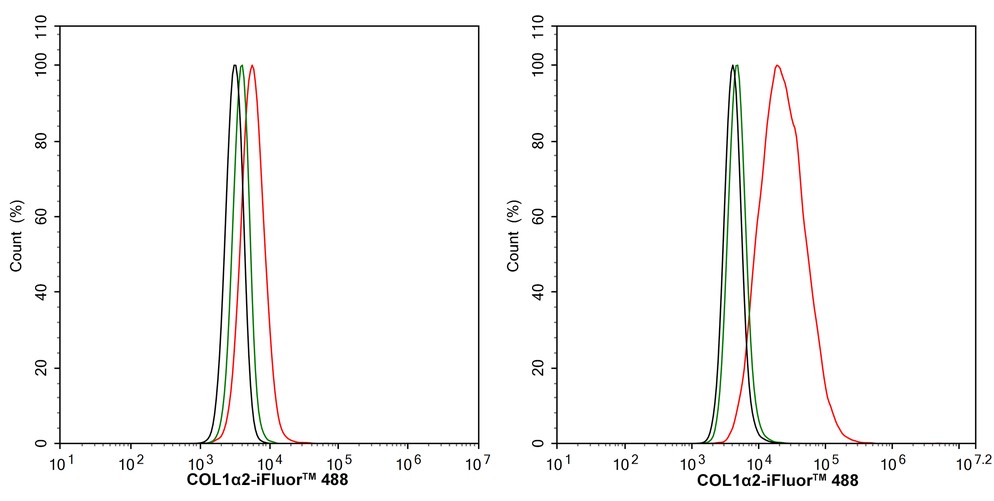
FC
Flow cytometric analysis of HeLa (left, negative) and T98G (right, positive) cells labeling COL1A2. Cells were washed twice with cold PBS and resuspend. Then stained with the primary antibody (1/1,000) (red) compared with Rabbit IgG Isotype Control (green). After incubation of the primary antibody at +4℃ for an hour, the cells were stained with a iFluor™ 488 conjugate-Goat anti-Rabbit IgG Secondary antibody at 1/1,000 dilution for 30 minutes at +4℃. Unlabelled sample was used as a control (cells without incubation with primary antibody; black).| Application Notes | WB:1:2000 ICC:1:100 FC:1:1000 |
|---|
| Form | Liquid |
|---|---|
| Storage Instructions | Store at +4℃ after thawing. Aliquot store at -20℃. Avoid repeated freeze / thaw cycles. |
| Storage Buffer | PBS (pH7.4), 0.1% BSA, 40% Glycerol. Preservative: 0.05% Sodium Azide. |
抱歉,暂无相关文献







