| 存储条件 |
|---|
| Product Name | GRP78 BiP Polyclonal Antibody |
|---|---|
| Antibody Type | Primary Antibodies |
| Immunogen | Synthetic peptide from human protein at AA range: 505-570 |
| Clonality | Polyclonal |
|---|---|
| Isotype | IgG |
| Host Species | Rabbit |
| Tested Applications | ELISAICC/IFIHCWB |
| WB:1:500-1:2000 IHC:1:50-1:300 ICC:1:50-1:200 | |
| Species Reactivity | FishHumanMouseRat |
| Concentration | 1mg/ml |
| Purification | Affinity purified |
| Gene Symbol | HSPA5 |
|---|---|
| Gene Synonyms | BIP GRP78 HEL-S-89n |
| Gene Full Name | heat shock protein family A (Hsp70) member 5 |
| Gene Summary | The protein encoded by this gene is a member of the heat shock protein 70 (HSP70) family. This protein localizes to the lumen of the endoplasmic reticulum (ER) where it operates as a typical HSP70 chaperone involved in the folding and assembly of proteins in the ER and is a master regulator of ER homeostasis. During cellular stress, as during viral infection or tumorogenesis, this protein interacts with the transmembrane stress sensor proteins PERK (protein kinase R-like endoplasmic reticulum kinase), IRE1 (inositol-requiring kinase 1), and ATF6 (activating transcription factor 6) where it acts as a repressor of the unfolded protein response (UPR) and also plays a role in cellular apoptosis and senescence. Elevated expression and atypical translocation of this protein to the cell surface has been reported in viral infections and some types of cancer cells. At the cell surface this protein may facilitate viral attachment and entry to host cells. This gene is a therapeutic target for the treatment of coronavirus diseases and chemoresistant cancers. [provided by RefSeq, Jul 2020] |
| Molecular Weight(MW) | 78kDa |
| Cellular Localization | Endoplasmic reticulum lumen . Melanosome . Cytoplasm . Cell surface . Identified by mass spectrometry in melanosome fractions from stage I to stage IV (PubMed:12643545). Localizes to the cell surface of epithelial cells in response to high levels of free iron (PubMed:20484814, PubMed:24355926, PubMed:27159390). . |
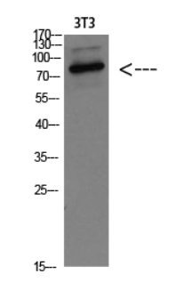
WB
Western blot analysis of 3T3 Cell Lysate, antibody was diluted at 1:1000. Secondary antibody was diluted at 1:20000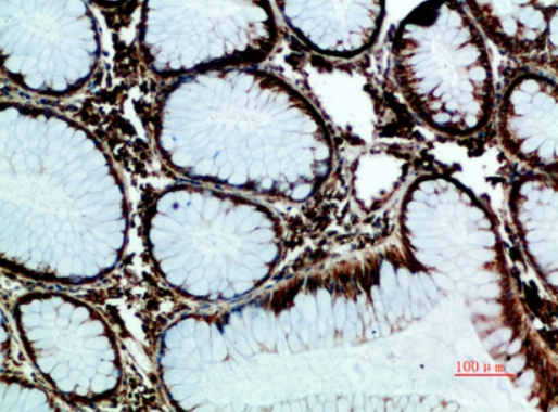
IHC
Immunohistochemical analysis of paraffin-embedded human-colon-cancer, antibody was diluted at 1:200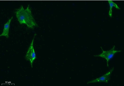
ICC/IF
Immunofluorescence analysis of A549. 1,primary Antibody was diluted at 1:200(4°C overnight). 2, Goat Anti Rabbit IgG (H&L) - Alexa Fluor 488 Secondary antibody was diluted at 1:1000(room temperature, 50min).3, Picture B: DAPI(blue) 10min.| Application Notes | WB:1:500-1:2000 IHC:1:50-1:300 ICC:1:50-1:200 |
|---|
| Form | Liquid |
|---|---|
| Storage Instructions | -15°C to -25°C/1 year(Do not lower than -25°C) |
| Storage Buffer | Liquid in PBS containing 50% glycerol, 0.5% BSA and 0.02% sodium azide. |
抱歉,暂无相关文献







