| 存储条件 |
|---|
| Product Name | TMEM119 Rabbit Polyclonal Antibody |
|---|---|
| Antibody Type | Primary Antibodies |
| Immunogen | Synthetic peptide within human TMEM119 aa 11-52 / 283. |
| Clonality | Polyclonal |
|---|---|
| Isotype | IgG |
| Host Species | Rabbit |
| Tested Applications | FCICC/IFWB |
| WB:1:5000 IF:1:50-1:100 FC:1:100-1:500 | |
| Species Reactivity | HumanMouse |
| Concentration | 1mg/ml |
| Purification | Affinity purified |
| Gene Symbol | TMEM119 |
|---|---|
| Gene Synonyms | OBIF |
| Molecular Weight(MW) | 29KD |
| Function | Plays an important role in bone formation and normal bone mineralization. Promotes the differentiation of myoblasts into osteoblasts. May induce the commitment and differentiation of myoblasts into osteoblasts through an enhancement of BMP2 production and interaction with the BMP-RUNX2 pathway. Upregulates the expression of ATF4, a transcription factor which plays a central role in osteoblast differentiation. Essential for normal spermatogenesis and late testicular differentiation. |
| Cellular Localization | Endoplasmic reticulum membrane, Cell membrane, Cytoplasm. |
| Protein Accession | Q4V9L6 |
|---|
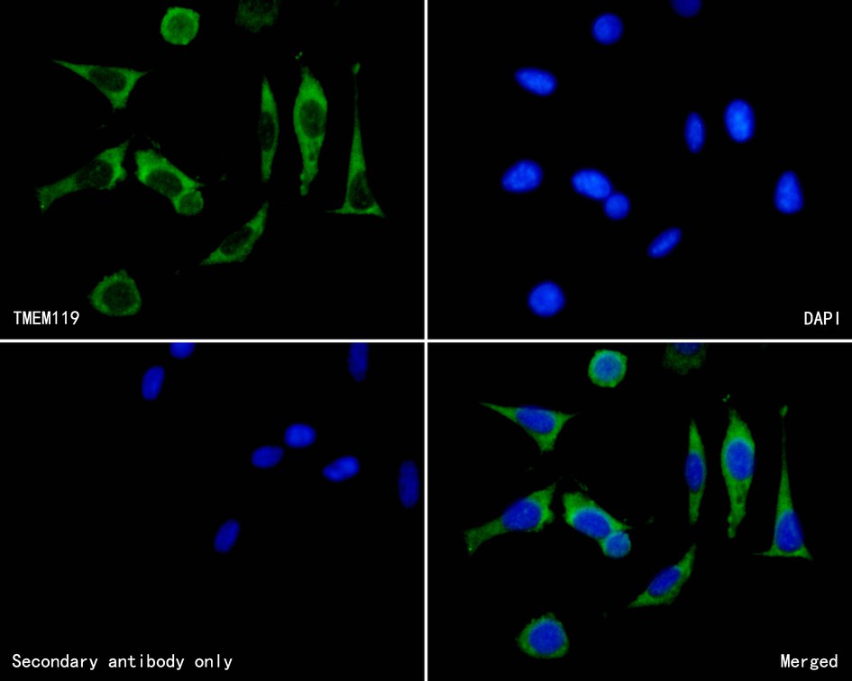
ICC/IF
ICC staining of TMEM119 in SH-SY5Y cells (green). Formalin fixed cells were permeabilized with 0.1% Triton X-100 in TBS for 10 minutes at room temperature and blocked with 10% negative goat serum for 15 minutes at room temperature. Cells were probed with the primary antibody (1/50) for 1 hour at room temperature, washed with PBS. Alexa Fluor®488 conjugate-Goat anti-Rabbit IgG was used as the secondary antibody at 1/1,000 dilution. The nuclear counter stain is DAPI (blue).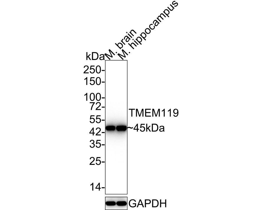
WB
Western blot analysis of TMEM119 on different lysates with Rabbit anti-TMEM119 antibody at 1/5,000 dilution. Lane 1: Mouse brain tissue lysate Lane 2: Mouse hippocampus tissue lysate Lysates/proteins at 20 µg/Lane. Predicted band size: 29 kDa Observed band size: 45 kDa Exposure time: 24 seconds; 4-20% SDS-PAGE gel. Proteins were transferred to a PVDF membrane and blocked with 5% NFDM/TBST for 1 hour at room temperature. The primary antibody at 1/5,000 dilution was used in 5% NFDM/TBST at 4℃ overnight. Goat Anti-Rabbit IgG - HRP Secondary Antibody at 1/50,000 dilution was used for 1 hour at room temperature.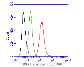
FC
Flow cytometric analysis of TMEM119 was done on SH-SY5Y cells. The cells were fixed, permeabilized and stained with the primary antibody (1ug/ml) (red) compared with Rabbit IgG, monoclonal - Isotype Control (green). After incubation of the primary antibody at +4℃ for 1 hour, the cells were stained with a Alexa Fluor®488 conjugate-Goat anti-Rabbit IgG Secondary antibody at 1/1,000 dilution for 30 minutes at +4℃ (dark incubation).Unlabelled sample was used as a control (cells without incubation with primary antibody; black).| Application Notes | WB:1:5000 IF:1:50-1:100 FC:1:100-1:500 |
|---|
| Form | Liquid |
|---|---|
| Storage Instructions | Store at +4℃ after thawing. Aliquot store at -20℃. Avoid repeated freeze / thaw cycles. |
| Storage Buffer | 1*TBS (pH7.4), 0.2% BSA, 50% Glycerol. Preservative: 0.05% Sodium Azide. |
抱歉,暂无相关文献







