| 存储条件 |
|---|
| Product Name | SDF1 |
|---|---|
| Antibody Type | Primary Antibodies |
| Antigen Alias | 12-O-tetradecanoylphorbol 13-acetate repressed protein 1 antibody AI174028 antibody C-X-C motif chemokine 12 antibody Chemokine (C-X-C motif) ligand 12 (stromal cell-derived factor 1) antibody Chemokine (C-X-C motif) ligand 12 antibody Chemokine CXC motif ligand 12 antibody cxcl12 antibody hIRH antibody hSDF-1 antibody Intercrine reduced in hepatomas antibody IRH antibody OTTHUMP00000019491 antibody PBSF antibody Pre-B cell growth-stimulating factor antibody SCYB12 antibody SDF 1 antibody SDF-1 antibody SDF-1-alpha(3-67) antibody SDF-1a antibody SDF-1b antibody SDF1_HUMAN antibody SDF1A antibody SDF1B antibody Stromal cell-derived factor 1 antibody Stromal cell-derived factor 1 delta antibody Stromal cell-derived factor 1 gamma antibody Stromal cell-derived factor 1a antibody Stromal cell-derived factor-1 alpha antibody Thymic lymphoma cell-stimulating factor antibody Tlsf antibody TLSF-a antibody TLSF-b antibody Tlsfa antibody Tlsfb antibody TPAR1 antibody |
| Product description | The C-X-C or α chemokine family is characterized by a pair of cysteine resi- dues separated by a single amino acid and primarily functions as chemoat- tractants for neutrophils. The C-X-C family includes IL-8, NAP-2, MSGA and stromal cell-derived factor-1, or SDF-1. SDF-1 was originally described as a pre-B cell stimulatory factor , but has now been shown to function as a potent chemoattractant for T cells and monocytes, but not neutrophils. Receptors for the C-X-C family are G protein-coupled, seven- pass, transmembrane do- main proteins which include IL-8RA, IL-8RB and fusin (also designated LESTR or CXCR-4). Fusin is highly homologous to the IL-8 receptors, sharing 37% sequence identity at the amino acid level. The IL-8 receptors bind to IL-8, NAP-2 and MSGA, while fusin binds to its cognate ligand, SDF-1. Fusin has been identified as the major co-receptor for T-tropic HIV-1, and SDF-1 has been shown to inhibit HIV-1 infection. Six human SDF-1 isoforms exist due to alternative splicing of CXCL12, the gene encoding SDF-1. Three isoforms are known for mouse and rat. |
| Immunogen | Amino acids 1-93 representing full-length SDF-1 of human origin |
| Clonality | Polyclonal |
|---|---|
| Isotype | IgG |
| Host Species | Rabbit |
| Tested Applications | WBIPIFIHC-P |
| WB:1:100-1:1,000 IP:1-2 μg per 100-500 μg of total protein (1 ml of cell lysate) IF:1:50-1:500 IHC:1:50-1:500 | |
| Species Reactivity | HumanMouseRat |
| Concentration | 1mg/ml |
| Alternative Names | 12-O-tetradecanoylphorbol 13-acetate repressed protein 1 antibody AI174028 antibody C-X-C motif chemokine 12 antibody Chemokine (C-X-C motif) ligand 12 (stromal cell-derived factor 1) antibody Chemokine (C-X-C motif) ligand 12 antibody Chemokine CXC motif ligand 12 antibody cxcl12 antibody hIRH antibody hSDF-1 antibody Intercrine reduced in hepatomas antibody IRH antibody OTTHUMP00000019491 antibody PBSF antibody Pre-B cell growth-stimulating factor antibody SCYB12 antibody SDF 1 antibody SDF-1 antibody SDF-1-alpha(3-67) antibody SDF-1a antibody SDF-1b antibody SDF1_HUMAN antibody SDF1A antibody SDF1B antibody Stromal cell-derived factor 1 antibody Stromal cell-derived factor 1 delta antibody Stromal cell-derived factor 1 gamma antibody Stromal cell-derived factor 1a antibody Stromal cell-derived factor-1 alpha antibody Thymic lymphoma cell-stimulating factor antibody Tlsf antibody TLSF-a antibody TLSF-b antibody Tlsfa antibody Tlsfb antibody TPAR1 antibody |
|---|---|
| Molecular Weight(MW) | 10kDa |
| Cellular Localization | Secreted |
| SwissProt ID | P48061 |
|---|
Application
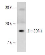
Fig1: Western blot analysis of SDF-1 expression in RAW264.7 whole cell lysate.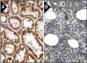
Application
Fig2: Immunoperoxidase staining of formalin fixed, paraffin-embedded human kidney tissue showing cytoplasmic staining of cells in tubules (A). Immunoperoxidase staining of formalin fixed, paraffin-embedded human bone marrow tissue showing nuclear and cytoplasmic staining of bone marrow poietic cells. (B).| Positive Control | RAW264.7, human kidney tissue, human bone marrow tissue. |
|---|---|
| Application Notes | WB:1:100-1:1,000 IP:1-2 μg per 100-500 μg of total protein (1 ml of cell lysate) IF:1:50-1:500 IHC:1:50-1:500 |
| Form | Liquid |
|---|---|
| Storage Instructions | Store at +4℃ |
| Storage Buffer | 1*TBS (pH7.4), 1%BSA, 40%Glycerol. Preservative: 0.05% Sodium Azide. |
抱歉,暂无相关文献
抱歉,暂无浏览记录







