| 存储条件 |
|---|
| Product Name | Histone H3 (tri methyl K9) Mouse Monoclonal Antibody |
|---|---|
| Antibody Type | Primary Antibodies |
| Product description | Eukaryotic histones are basic and water soluble nuclear proteins that form hetero-octameric nucleosome particles by wrapping 146 base pairs of DNA in a left-handed super-helical turn sequentially to form chromosomal fibers. Two molecules of each of the four core histones (H2A, H2B, H3 and H4) form the octamer, which is comprised of two H2A-H2B dimers and two H3-H4 dimers, forming two nearly symmetrical halves by tertiary structure. Histones are subject to posttranslational modification by enzymes primarily on their N-terminal tails, but also in their globular domains. Human Histone H3 is subject to trimethylation at Lys 9, a modification that may be necessary for select DNA transactions or chromatin state transitions. Acetylation is generally linked to gene activation. Acetylation on Lys-10 (H3K9ac) impairs methylation at Arg-9 (H3R8me2s). Acetylation on Lys-19 (H3K18ac) and Lys-24 (H3K24ac) favors methylation at Arg-18 (H3R17me). Citrullination at Arg-9 (H3R8ci) and/or Arg-18 (H3R17ci) by PADI4 impairs methylation and represses transcription. Asymmetric dimethylation at Arg-18 (H3R17me2a) by CARM1 is linked to gene activation. Symmetric dimethylation at Arg-9 (H3R8me2s) by PRMT5 is linked to gene repression. Asymmetric dimethylation at Arg-3 (H3R2me2a) by PRMT6 is linked to gene repression and is mutually exclusive with H3 Lys-5 methylation (H3K4me2 and H3K4me3). H3R2me2a is present at the 3' of genes regardless of their transcription state and is enriched on inactive promoters, while it is absent on active promoters. |
| Immunogen | Synthetic peptide within N terminal human Histone H3 including tri methyl K9 conjugated to keyhole limpet haemocyanin. |
| Modification | tri methyl K9 |
| Clonality | Monoclonal |
|---|---|
| Isotype | IgG1 |
| Host Species | Mouse |
| Tested Applications | ICC/IFIHCWB |
| WB:1:1000-1:5000 IHC:1:1000 ICC/IF:1:250 | |
| Species Reactivity | HumanMouseRat |
| Concentration | 1mg/ml |
| Purification | Protein G |
| Alternative Names | H3 histone family member E pseudogene antibody H3 histone family member A antibody H3/A antibody H31_HUMAN antibody H3F3 antibody H3FA antibody Hist1h3a antibody HIST1H3B antibody HIST1H3C antibody HIST1H3D antibody HIST1H3E antibody HIST1H3F antibody HIST1H3G antibody HIST1H3H antibody HIST1H3I antibody HIST1H3J antibody HIST3H3 antibody histone 1 H3a antibody Histone cluster 1 H3a antibody Histone H3 3 pseudogene antibody Histone H3.1 antibody Histone H3/a antibody Histone H3/b antibody Histone H3/c antibody Histone H3/d antibody Histone H3/f antibody Histone H3/h antibody Histone H3/i antibody Histone H3/j antibody Histone H3/k antibody Histone H3/l antibody |
|---|---|
| Molecular Weight(MW) | 15kDa |
| Cellular Localization | Nucleus. |
| SwissProt ID | P68431 |
|---|
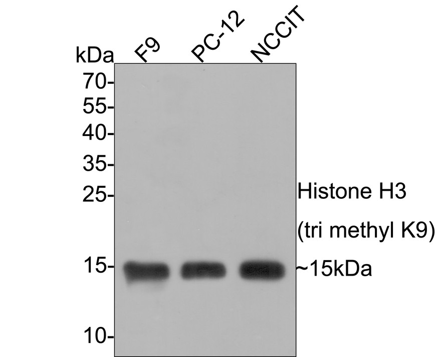
WB
Western blot analysis of Histone H3 (tri methyl K9) on different lysates with Mouse anti-Histone H3 (tri methyl K9) antibody at 1/1,000 dilution. Lane 1: F9 cell lysate, Lane 2: PC-12 cell lysate, Lane 3: NCCIT cell lysate, Lysates/proteins at 10 µg/Lane. Exposure time: minute; 15% SDS-PAGE gel. Proteins were transferred to a PVDF membrane and blocked with 5% NFDM/TBST for 1 hour at room temperature. The primary antibody at 1/1,000 dilution was used in 5% NFDM/TBST at room temperature for 2 hours. Goat Anti-Mouse IgG - HRP Secondary Antibody at 1:100,000 dilution was used for 1 hour at room temperature.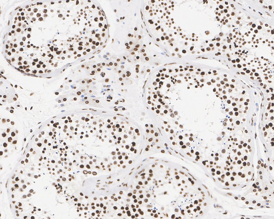
IHC
Immunohistochemical analysis of paraffin-embedded human testis tissue with Mouse anti-Histone H3 (tri methyl K9) antibody at 1/1,000 dilution. The section was pre-treated using heat mediated antigen retrieval with sodium citrate buffer (pH 6.0) (high pressure) for 2 minutes. The tissues were blocked in 1% BSA for 20 minutes at room temperature, washed with ddH2O and PBS, and then probed with the primary antibody at 1/1,000 dilution for 1 hour at room temperature. The detection was performed using an HRP conjugated compact polymer system. DAB was used as the chromogen. Tissues were counterstained with hematoxylin and mounted with DPX.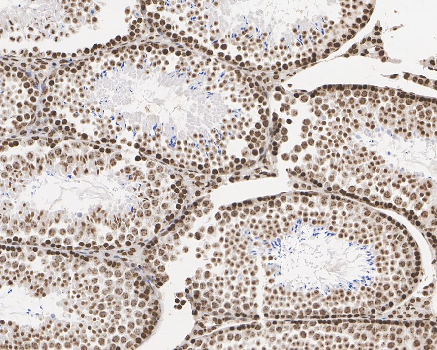
IHC
Immunohistochemical analysis of paraffin-embedded mouse testis tissue with Mouse anti-Histone H3 (tri methyl K9) antibody at 1/1,000 dilution. The section was pre-treated using heat mediated antigen retrieval with sodium citrate buffer (pH 6.0) (high pressure) for 2 minutes. The tissues were blocked in 1% BSA for 20 minutes at room temperature, washed with ddH2O and PBS, and then probed with the primary antibody at 1/1,000 dilution for 1 hour at room temperature. The detection was performed using an HRP conjugated compact polymer system. DAB was used as the chromogen. Tissues were counterstained with hematoxylin and mounted with DPX.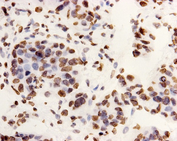
IHC
Immunohistochemical analysis of paraffin-embedded rat testis tissue using anti-Tri-Methyl-Histone H3 (Lys9) antibody. The section was pre-treated using heat mediated antigen retrieval with Tris-EDTA buffer (pH 8.0-8.4) for 20 minutes.The tissues were blocked in 5% BSA for 30 minutes at room temperature, washed with ddH2O and PBS, and then probed with the primary antibody (1/100) for 30 minutes at room temperature. The detection was performed using an HRP conjugated compact polymer system. DAB was used as the chromogen. Tissues were counterstained with hematoxylin and mounted with DPX.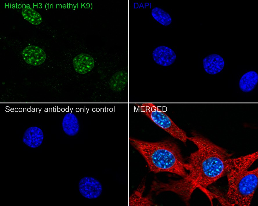
ICC/IF
Immunocytochemistry analysis of NIH/3T3 cells labeling Histone H3 (tri methyl K9) with Mouse anti-Histone H3 (tri methyl K9) antibody at 1/250 dilution. Cells were fixed in 4% paraformaldehyde for 20 minutes at room temperature, permeabilized with 0.1% Triton X-100 in PBS for 5 minutes at room temperature, then blocked with 1% BSA in 10% negative goat serum for 1 hour at room temperature. Cells were then incubated with Mouse anti-Histone H3 (tri methyl K9) antibody at 1/250 dilution in 1% BSA in PBST overnight at 4 ℃. Goat Anti-Mouse IgG H&L (488) was used as the secondary antibody at 1/1,000 dilution. PBS instead of the primary antibody was used as the secondary antibody only control. Nuclear DNA was labelled in blue with DAPI. beta Tubulin (red) was stained at 1/100 dilution overnight at +4℃. Goat Anti-Rabbit IgG H&L (594) were used as the secondary antibody at 1/1,000 dilution.| Application Notes | WB:1:1000-1:5000 IHC:1:1000 ICC/IF:1:250 |
|---|
| Form | Liquid |
|---|---|
| Storage Instructions | Store at +4℃ short term (1-2 weeks). It is recommended to aliquot into single-use upon delivery. Store at -20℃ long term. |
| Storage Buffer | 1*PBS (pH7.4), 0.2% BSA, 50% Glycerol. Preservative: 0.05% Sodium Azide. |
抱歉,暂无相关文献
抱歉,暂无浏览记录







