| 存储条件 |
|---|
| Product Name | MTERFD1 |
|---|---|
| Antibody Type | Primary Antibodies |
| Antigen Alias | CGI 12 antibody mitochondrial antibody Mitochondrial transcription termination factor 3 antibody MTER1_HUMAN antibody MTERF domain containing 1 antibody mTERF domain containing protein 1 mitochondrial antibody mTERF domain-containing protein 1 antibody mTERF3 antibody Mterfd1 antibody Transcription termination factor 3, mitochondrial antibody |
| Product description | Binds promoter DNA and regulates initiation of transcription. Required for normal mitochondrial transcription and translation, and for normal assembly of mitochondrial respiratory complexes. Required for normal mitochondrial function.Maintains 16S rRNA levels and functions in mitochondrial ribosome assembly by regulating the biogenesis of the 39S ribosomal subunit. |
| Immunogen | Recombinant protein. |
| Clonality | Polyclonal |
|---|---|
| Isotype | IgG |
| Host Species | Rabbit |
| Tested Applications | WBICCIHC |
| WB:1:500~1:2000 ICC:1:200~1:500 IHC:1:50~1:200 Notes:Optimal dilutions/concentrations should be determined by the researcher. | |
| Species Reactivity | HumanMouseRat |
| Concentration | 1mg/ml |
| Alternative Names | CGI 12 antibody mitochondrial antibody Mitochondrial transcription termination factor 3 antibody MTER1_HUMAN antibody MTERF domain containing 1 antibody mTERF domain containing protein 1 mitochondrial antibody mTERF domain-containing protein 1 antibody mTERF3 antibody Mterfd1 antibody Transcription termination factor 3 mitochondrial antibody |
|---|---|
| Molecular Weight(MW) | 38 kDa |
| Cellular Localization | Mitochondrion. |
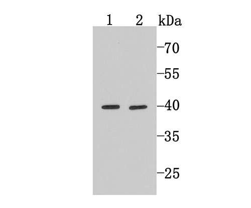
Application
Fig1: Western blot analysis of MTERFD1 on Hela (1) and HepG2 (2) cell lysates using anti-MTERFD1 antibody at 1/500 dilution.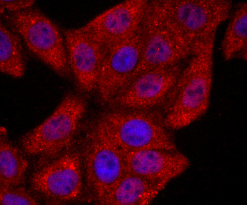
Application
Fig2: ICC staining MTERFD1 in HepG2 cells (red). The nuclear counter stain is DAPI (blue). Cells were fixed in paraformaldehyde, permeabilised with 0.25% Triton X100/PBS.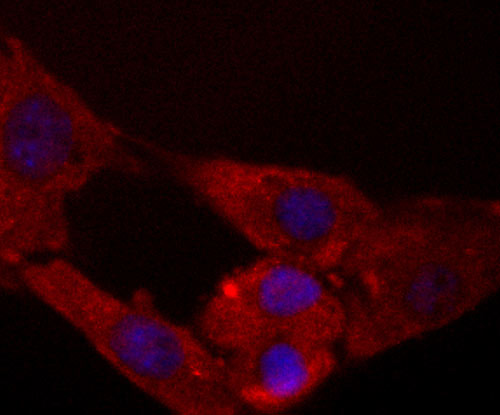
Application
Fig3: ICC staining MTERFD1 in NIH-3T3 cells (red). The nuclear counter stain is DAPI (blue). Cells were fixed in paraformaldehyde, permeabilised with 0.25% Triton X100/PBS.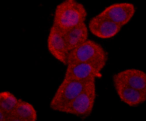
Application
Fig4: ICC staining MTERFD1 in Hela cells (red). The nuclear counter stain is DAPI (blue). Cells were fixed in paraformaldehyde, permeabilised with 0.25% Triton X100/PBS.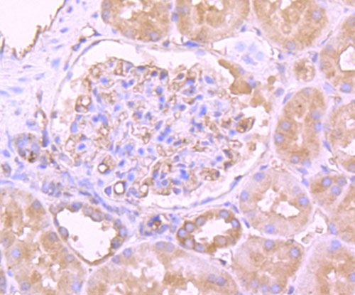
Application
Fig5: Immunohistochemical analysis of paraffin-embedded human kidney tissue using anti-MTERFD1 antibody. Counter stained with hematoxylin.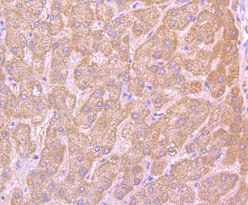
Application
Fig6: Immunohistochemical analysis of paraffin-embedded human liver tissue using anti-MTERFD1 antibody. Counter stained with hematoxylin.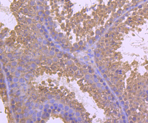
Application
Fig7: Immunohistochemical analysis of paraffin-embedded mouse testis tissue using anti-MTERFD1 antibody. Counter stained with hematoxylin.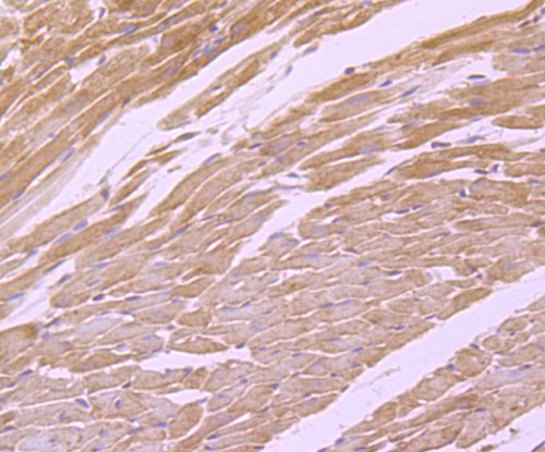
Application
Fig8: Immunohistochemical analysis of paraffin-embedded mouse heart tissue using anti-MTERFD1 antibody. Counter stained with hematoxylin.| Positive Control | Hela, HepG2, NIH-3T3, human liver tissue, human kidney tissue, mouse testis tissue, mouse heart tissue. |
|---|---|
| Application Notes | WB:1:500~1:2000 ICC:1:200~1:500 IHC:1:50~1:200 Notes:Optimal dilutions/concentrations should be determined by the researcher. |
| Form | Liquid |
|---|---|
| Storage Instructions | Store at +4℃ after thawing. Aliquot store at -20℃ or -80℃. Avoid repeated freeze / thaw cycles. |
| Storage Buffer | 1*TBS (pH7.4), 0.5%BSA, 50%Glycerol. Preservative: 0.05% Sodium Azide. |
抱歉,暂无相关文献
抱歉,暂无浏览记录







