| 存储条件 |
|---|
| Product Name | GPC1 |
|---|---|
| Antibody Type | Primary Antibodies |
| Antigen Alias | FLJ38078 antibody glypican antibody Glypican proteoglycan 1 antibody glypican1 antibody GPC 1 antibody Gpc1 antibody GPC1_HUMAN antibody Secreted glypican-1 antibody |
| Product description | Glypican-1 (GPC1), a member of the glycosylphosphatidilinositol anchored cell surface heparan sulfate proteoglycans, is involved with cell adhesion and migration, lipoprotein metabolism, modulation of growth factor activites and anticoagulation. Glypican-1 binds to and modulates the activity of several fibroblast growth factors (FGFs) including FGF-1, FGF-2 and FGF-7. Glypican-1 acts as an extracellular chaperone for VEGF165 to help restore receptor binding ability after oxidation. The heparan sulfate chains of glypican-1 mediate specific binding of glypican-1 to VEGF165. When present on the surface of marrow stromal cells, glypican-1 may aid in the maintenance and development of hematopoietic stem and progenitor cells. Human pancreatic cancer cells express a large amount of glypican-1 when compared to glypican-1 levels in normal pancreatic cells. Glypican-1 may play an important role in the response of pancreatic cancer cells to mitogenic stimuli, such as FGF-2. The gene encoding human glypican-1 maps to chromosome 2q37.3. |
| Immunogen | Peptide |
| Clonality | Polyclonal |
|---|---|
| Isotype | IgG |
| Host Species | Rabbit |
| Tested Applications | WBICCIHCFC |
| WB:1:500-1:1,000 ICC:1:50-1:200 IHC:1:50-1:200 FC:1:50-1:100 | |
| Species Reactivity | Human |
| Concentration | 1mg/ml |
| Alternative Names | FLJ38078 antibody glypican antibody Glypican proteoglycan 1 antibody glypican1 antibody GPC 1 antibody Gpc1 antibody GPC1_HUMAN antibody Secreted glypican-1 antibody |
|---|---|
| Molecular Weight(MW) | 61 kDa |
| Cellular Localization | Membrane. Secreted. |
| SwissProt ID | P35052 |
|---|
Application
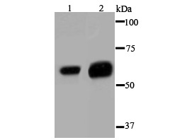
Fig1: Western blot analysis of Glypican-1 on different cell lysate using anti- Glypican-1 antibody at 1/1,000 dilution. Positive control: Lane1: Hela Lane2: SK-Br-3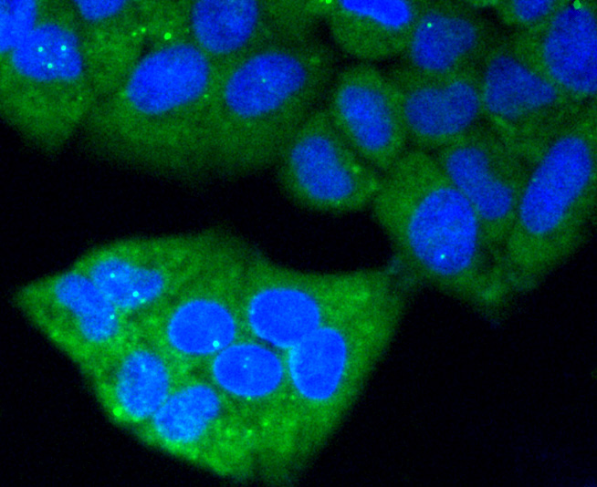
Application
Fig2: ICC staining Glypican-1 in Hela cells (green). The nuclear counter stain is DAPI (blue). Cells were fixed in paraformaldehyde, permeabilised with 0.25% Triton X100/PBS.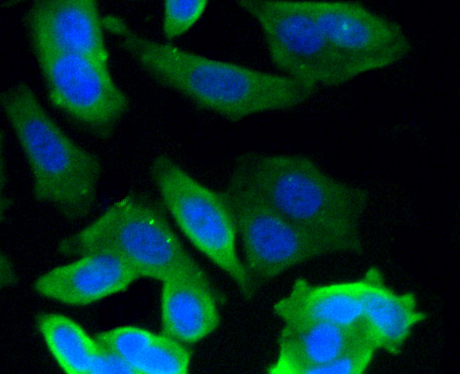
Application
Fig3: ICC staining Glypican-1 in MCF-7 cells (green). The nuclear counter stain is DAPI (blue). Cells were fixed in paraformaldehyde, permeabilised with 0.25% Triton X100/PBS.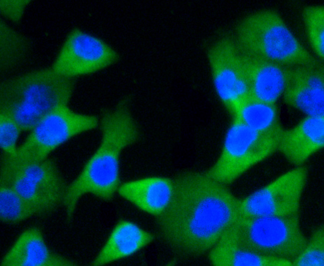
Application
Fig4: ICC staining Glypican-1 in PANC-1 cells (green). The nuclear counter stain is DAPI (blue). Cells were fixed in paraformaldehyde, permeabilised with 0.25% Triton X100/PBS.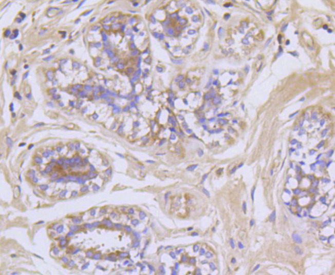
Application
Fig5: Immunohistochemical analysis of paraffin-embedded human breast tissue using anti- Glypican-1 antibody. Counter stained with hematoxylin.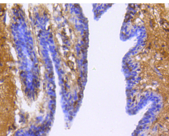
Application
Fig6: Immunohistochemical analysis of paraffin-embedded human breast cancer tissue using anti- Glypican-1 antibody. Counter stained with hematoxylin.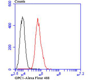
Application
Fig7: Flow cytometric analysis of MCF-7 cells with Glypican-1 antibody at 1/100 dilution (red) compared with an unlabelled control (cells without incubation with primary antibody; black).| Positive Control | Hela, SK-Br-3, MCF-7, PANC-1, human breast tissue, human breast cancer tissue. |
|---|---|
| Application Notes | WB:1:500-1:1,000 ICC:1:50-1:200 IHC:1:50-1:200 FC:1:50-1:100 |
| Form | Liquid |
|---|---|
| Storage Instructions | Store at +4℃after thawing. Aliquot store at -20℃or -80℃. Avoid repeated freeze / thaw cycles. |
| Storage Buffer | 1*TBS (pH7.4), 0.5%BSA, 50%Glycerol. Preservative: 0.05% Sodium Azide. |
抱歉,暂无相关文献







