| 存储条件 |
|---|
| Product Name | FPR1 |
|---|---|
| Antibody Type | Primary Antibodies |
| Antigen Alias | fMet Leu Phe receptor antibody fMet-Leu-Phe receptor antibody FMLP antibody fMLP receptor antibody FMLPR antibody Formyl peptide receptor 1 antibody FPR 1 antibody FPR antibody FPR receptor antibody FPR1 antibody FPR1_HUMAN antibody N formyl peptide chemoattractant receptor antibody N formyl peptide receptor antibody N formylpeptide chemoattractant receptor antibody N-formyl peptide receptor antibody N-formylpeptide chemoattractant receptor antibody |
| Product description | High affinity receptor for N-formyl-methionyl peptides, which are powerful neutrophils chemotactic factors. Binding of FMLP to the receptor causes activation of neutrophils. This response is mediated via a G-protein that activates a phosphatidylinositol-calcium second messenger system. |
| Immunogen | Peptide |
| Clonality | Polyclonal |
|---|---|
| Isotype | IgG |
| Host Species | Rabbit |
| Tested Applications | WBICCIHCFC |
| WB:1:500 ICC:1:50-1:200 IHC:1:50-1:100 FC:1:50-1:100 | |
| Species Reactivity | HumanMouse |
| Concentration | 1mg/ml |
| Alternative Names | fMet Leu Phe receptor antibody fMet-Leu-Phe receptor antibody FMLP antibody fMLP receptor antibody FMLPR antibody Formyl peptide receptor 1 antibody FPR 1 antibody FPR antibody FPR receptor antibody FPR1 antibody FPR1_HUMAN antibody N formyl peptide chemoattractant receptor antibody N formyl peptide receptor antibody N formylpeptide chemoattractant receptor antibody N-formyl peptide receptor antibody N-formylpeptide chemoattractant receptor antibody |
|---|---|
| Molecular Weight(MW) | 38 kDa |
| Cellular Localization | Cell membrane. |
| SwissProt ID | P21462 |
|---|
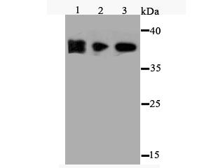
Application
at 1/500 dilution. Positive control: Lane1: NCCIT Lane2: MEF Lane3: HES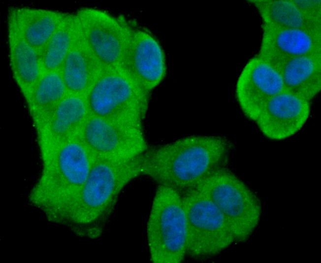
Application
Fig2: ICC staining FPR1 in Hela cells (green). The nuclear counter stain is DAPI (blue). Cells were fixed in paraformaldehyde, permeabilised with 0.25% Triton X100/PBS.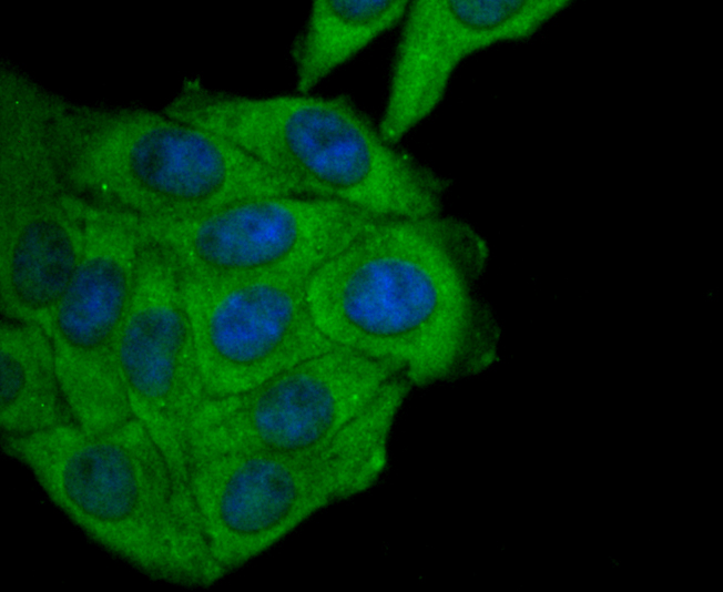
Application
Fig3: ICC staining FPR1 in HepG2 cells (green). The nuclear counter stain is DAPI (blue). Cells were fixed in paraformaldehyde, permeabilised with 0.25% Triton X100/PBS.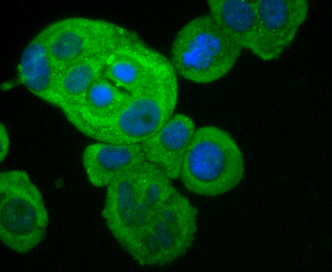
Application
Fig4: ICC staining FPR1 in MCF-7 cells (green). The nuclear counter stain is DAPI (blue). Cells were fixed in paraformaldehyde, permeabilised with 0.25% Triton X100/PBS.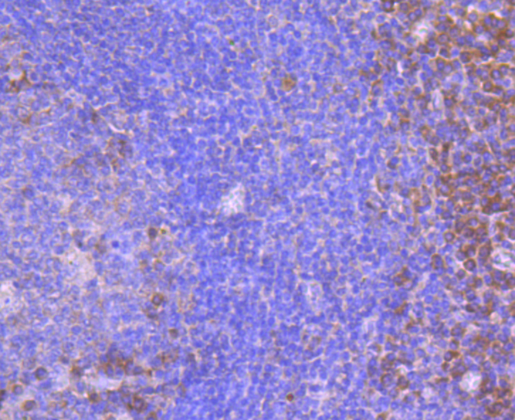
Application
Fig5: Immunohistochemical analysis of paraffin-embedded human tonsil tissue using anti-FPR1 antibody. Counter stained with hematoxylin.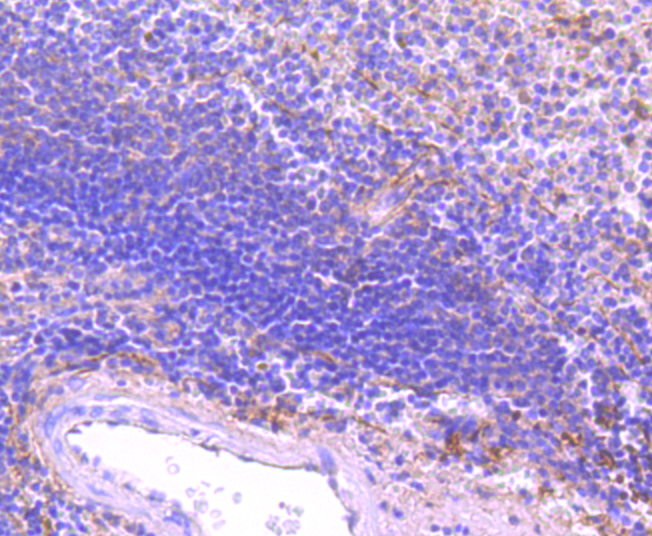
Application
Fig6: Immunohistochemical analysis of paraffin-embedded human spleen tissue using anti-FPR1 antibody. Counter stained with hematoxylin.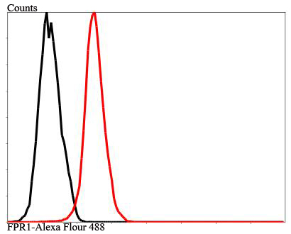
Application
Fig7: Flow cytometric analysis of MCF-7 cells with FPR1 antibody at 1/100 dilution (red) compared with an unlabelled control (cells without incubation with primary antibody; black).| Positive Control | NCCIT, MEF, HES, Hela, HepG2, MCF-7, human tonsil tissue, human spleen tissue. |
|---|---|
| Application Notes | WB:1:500 ICC:1:50-1:200 IHC:1:50-1:100 FC:1:50-1:100 |
| Form | Liquid |
|---|---|
| Storage Instructions | Store at +4℃ after thawing. Aliquot store at -20℃ or -80℃. Avoid repeated freeze / thaw cycles. |
| Storage Buffer | 1*TBS (pH7.4), 1%BSA, 40%Glycerol. Preservative: 0.05% Sodium Azide. |
抱歉,暂无相关文献







