| 存储条件 |
|---|
| Product Name | PHD2 |
|---|---|
| Antibody Type | Primary Antibodies |
| Antigen Alias | C1ORF12 antibody Chromosome 1 Open Reading Frame 12 antibody DKFZp761F179 antibody ECYT 3 antibody ECYT3 antibody EGL 9 homolog of C. elegans 1 antibody EGL nine (C.elegans) homolog 1 antibody Egl nine homolog 1 (C. elegans) antibody Egl nine homolog 1 antibody Egl nine like protein 1 antibody EGLN 1 antibody Egln1 antibody EGLN1_HUMAN antibody HIF PH2 antibody HIF Prolyl Hydroxylase 2 antibody HIF-PH2 antibody HIF-prolyl hydroxylase 2 antibody HIFP4H 2 antibody HIFPH2 antibody HPH 2 antibody HPH-2 antibody HPH2 antibody Hypoxia inducible factor prolyl hydroxylase 2 antibody Hypoxia-inducible factor prolyl hydroxylase 2 antibody ORF13 antibody P4H2 antibody PHD 2 antibody PhD2 antibody PNAS 118 antibody PNAS 137 antibody Prolyl Hydroxylase Domain Containing Protein 2 antibody Prolyl hydroxylase domain-containing protein 2 antibody Rat Homolog of SM20 antibody SM 20 antibody SM-20 antibody SM20 antibody Zinc finger MYND domain containing protein 6 antibody ZMYND6 antibody |
| Product description | Prolyl hydroxylase domain proteins HIF PHD1, HIF PHD2 and HIF PHD3 (known as PHD1, PHD2 and PHD3 in rodents, respectively) can hydroxylate HIF-α subunits. Hypoxia-inducible factor (HIF) is a transcriptional regulator important in several aspects of oxygen homeostasis. The prolyl hydroxylases catalyze the posttranslational formation of 4-hydroxyproline in HIF-α proteins. HIF PHD1, which is widely expressed, with highest levels of expression in testis, functions as a cellular oxygen sensor and is important in cell growth regulation. HIF PHD1 can localize to the nucleus or the cytoplasm and is also detected in hormone responsive tissues, such as normal and cancerous mammary, ovarian and prostate epithelium. HIF PHD1 is encoded by EGLN2, which maps to chromosome 19q13.3. HIF PHD2 is regarded as the main cellular oxygen sensor, as RNA interference against HIF PHD2, but not HIF PHD1 or HIF PHD3, is enough to stabilize HIF-1α in normoxia. HIF PHD2, a direct HIF target gene, is expressed mainly in skeletal muscle, heart, kidney and brain. |
| Immunogen | peptide |
| Clonality | Polyclonal |
|---|---|
| Isotype | IgG |
| Host Species | Rabbit |
| Tested Applications | WBICCIHCFC |
| WB:1:1,000 ICC:1:50-1:200 IHC:1:50-1:200 FC:1:10-1:100 | |
| Species Reactivity | HumanMouseRat |
| Concentration | 1mg/ml |
| Alternative Names | C1ORF12 antibody Chromosome 1 Open Reading Frame 12 antibody DKFZp761F179 antibody ECYT 3 antibody ECYT3 antibody EGL 9 homolog of C. elegans 1 antibody EGL nine (C.elegans) homolog 1 antibody Egl nine homolog 1 (C. elegans) antibody Egl nine homolog 1 antibody Egl nine like protein 1 antibody EGLN 1 antibody Egln1 antibody EGLN1_HUMAN antibody HIF PH2 antibody HIF Prolyl Hydroxylase 2 antibody HIF-PH2 antibody HIF-prolyl hydroxylase 2 antibody HIFP4H 2 antibody HIFPH2 antibody HPH 2 antibody HPH-2 antibody HPH2 antibody Hypoxia inducible factor prolyl hydroxylase 2 antibody Hypoxia-inducible factor prolyl hydroxylase 2 antibody ORF13 antibody P4H2 antibody PHD 2 antibody PhD2 antibody PNAS 118 antibody PNAS 137 antibody Prolyl Hydroxylase Domain Containing Protein 2 antibody Prolyl hydroxylase domain-containing protein 2 antibody Rat Homolog of SM20 antibody SM 20 antibody SM-20 antibody SM20 antibody Zinc finger MYND domain containing protein 6 antibody ZMYND6 antibody |
|---|---|
| Molecular Weight(MW) | 46kDa |
| Cellular Localization | Cytoplasm, Nucleus |
| SwissProt ID | Q9GZT9 |
|---|
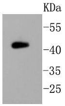
Application
Fig1: Western blot analysis on mouse brain lysates using anti-PHD2 rabbit polyclonal antibody.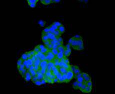
Application
Fig2: Immunocytochemical staining of PC-12 cells using anti-PHD2 rabbit polyclonal antibody.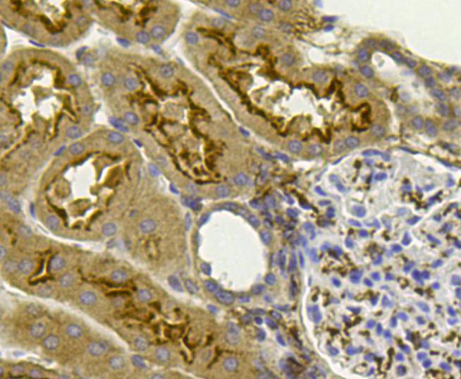
Application
Fig3: Immunohistochemical analysis of paraffin- embedded human kidney tissue using anti-PHD2 rabbit polyclonal antibody.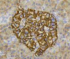
Application
Fig4: Immunohistochemical analysis of paraffin- embedded human pancreas tissue using anti-PHD2 rabbit polyclonal antibody.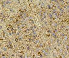
Application
Fig5: Immunohistochemical analysis of paraffin- embedded mouse brain tissue using anti-PHD2 rabbit polyclonal antibody.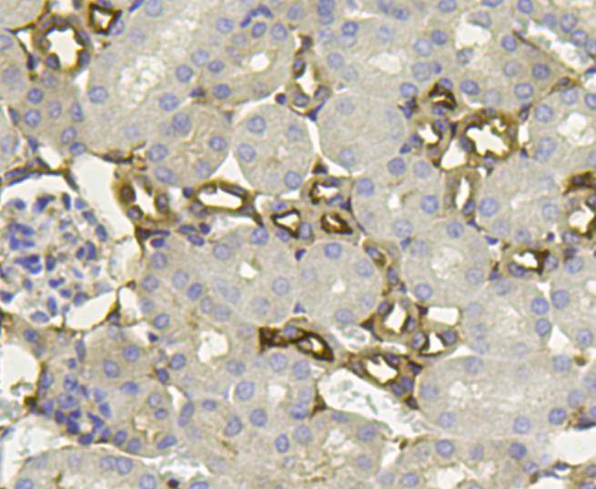
Application
Fig6: Immunohistochemical analysis of paraffin- embedded mouse kidney tissue using anti-PHD2 rabbit polyclonal antibody.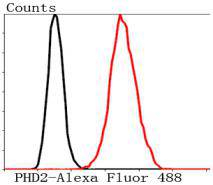
Application
Fig7: Flow cytometric analysis of SH-SY-5Y cells with PHD2 antibody at 1/50 dilution (blue) compared with an unlabelled control (cells without incubation with primary antibody; red). Alexa Fluor 488-conjugated Goat anti rabbit IgG was used as the secondary antibody.| Positive Control | PC-12, human kidney tissue, mouse kidney tissue, human pancreas tissue, mouse brain tissue. |
|---|---|
| Application Notes | WB:1:1,000 ICC:1:50-1:200 IHC:1:50-1:200 FC:1:10-1:100 |
| Form | Liquid |
|---|---|
| Storage Instructions | Store at +4℃ after thawing. Aliquot store at -20℃ or -80℃. Avoid repeated freeze / thaw cycles. |
| Storage Buffer | 1*TBS (pH7.4), 1%BSA, 40%Glycerol. Preservative: 0.05% Sodium Azide. |
抱歉,暂无相关文献







