| 存储条件 |
|---|
| Product Name | BMPR1A |
|---|---|
| Antibody Type | Primary Antibodies |
| Antigen Alias | 10q23del antibody Activin A receptor type II like kinase 3 antibody Activin receptor like kinase 3 antibody Activin receptor-like kinase 3 antibody ACVRLK 3 antibody ACVRLK3 antibody ALK 3 antibody ALK-3 antibody ALK3 antibody BMP type-1A receptor antibody BMPR 1A antibody Bmpr antibody BMPR-1A antibody Bmpr1a antibody BMR1A_HUMAN antibody Bone morphogenetic protein receptor type IA antibody Bone morphogenetic protein receptor type IA precursor antibody Bone morphogenetic protein receptor type-1A antibody BR 1a antibody BR1a antibody CD 292 antibody CD292 antibody CD292 antigen antibody EC 2.7.11.30 antibody Serine threonine protein kinase receptor R5 antibody Serine threonine protein kinase receptor R5 precursor antibody Serine/threonine-protein kinase receptor R5 antibody SKR 5 antibody SKR5 antibody zBMPR IA antibody zBMPRIA antibody |
| Product description | Members of the transforming growth factor b superfamily bind to a pair of transmembrane proteins, known as receptor types I and II, which contain serine/threonine kinases and associate to form a signaling complex. Two type I receptors have been characterized, BMPR-IA (also designated SKR5, ALK-3, and BRK-1) and BMPR-IB (also designated ALK-6 and SKR 6), that bind to bone morphogenetic proteins (BMP)-2, BMP-4, and osteogenic protein (OP)-1 (also designated BMP-7). BMPR-IA and BMPR-IB are both expressed in human glioma cell lines. The type II receptor, BMPR-II, efficiently binds to OP-1 and BMP-2 and weakly binds BMP-4, and it is widely expressed in different tissues, including brain. The BMP receptor family members are thought to mediate distinct effects on gene expression, cell differentiation, and morphogenesis in a dose dependent fashion. |
| Immunogen | recombinant protein |
| Clonality | Polyclonal |
|---|---|
| Isotype | IgG |
| Host Species | Rabbit |
| Tested Applications | WBICCIHCFC |
| WB:1:1,1000 ICC:1:50-1:200 IHC:1:50-1:200 FC:1:50-1:100 | |
| Species Reactivity | HumanMouseRat |
| Concentration | 1mg/ml |
| Alternative Names | 10q23del antibody Activin A receptor type II like kinase 3 antibody Activin receptor like kinase 3 antibody Activin receptor-like kinase 3 antibody ACVRLK 3 antibody ACVRLK3 antibody ALK 3 antibody ALK-3 antibody ALK3 antibody BMP type-1A receptor antibody BMPR 1A antibody Bmpr antibody BMPR-1A antibody Bmpr1a antibody BMR1A_HUMAN antibody Bone morphogenetic protein receptor type IA antibody Bone morphogenetic protein receptor type IA precursor antibody Bone morphogenetic protein receptor type-1A antibody BR 1a antibody BR1a antibody CD 292 antibody CD292 antibody CD292 antigen antibody EC 2.7.11.30 antibody Serine threonine protein kinase receptor R5 antibody Serine threonine protein kinase receptor R5 precursor antibody Serine/threonine-protein kinase receptor R5 antibody SKR 5 antibody SKR5 antibody zBMPR IA antibody zBMPRIA antibody |
|---|---|
| Molecular Weight(MW) | 60kDa |
| Cellular Localization | Membrane |
| SwissProt ID | P36894 |
|---|
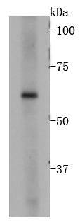
Application
Fig1: Western blot analysis on 293 cell lysates using anti-BMPR1A rabbit polyclonal antibody.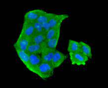
Application
Fig2: Immunocytochemical staining of Hela cells using anti-BMPR1A rabbit polyclonal antibody.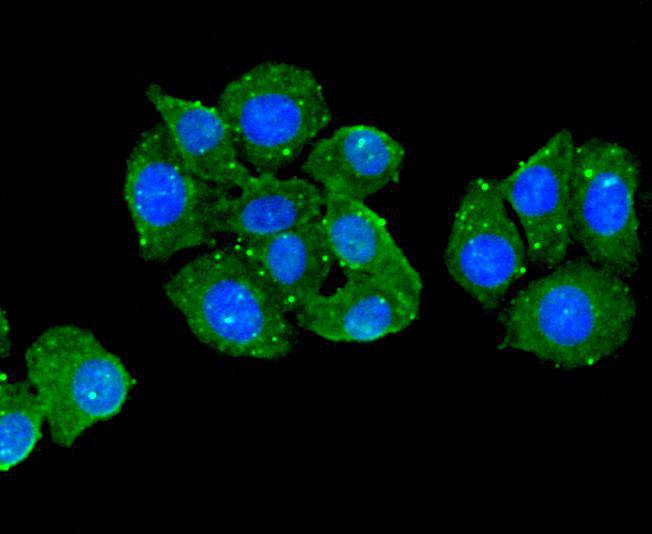
Application
Fig3: Immunocytochemical staining of HepG2 cells using anti-BMPR1A rabbit polyclonal antibody.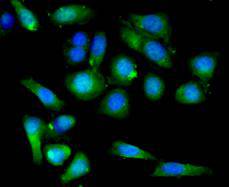
Application
Fig4: Immunocytochemical staining of SKOV-3 cells using anti-BMPR1A rabbit polyclonal antibody.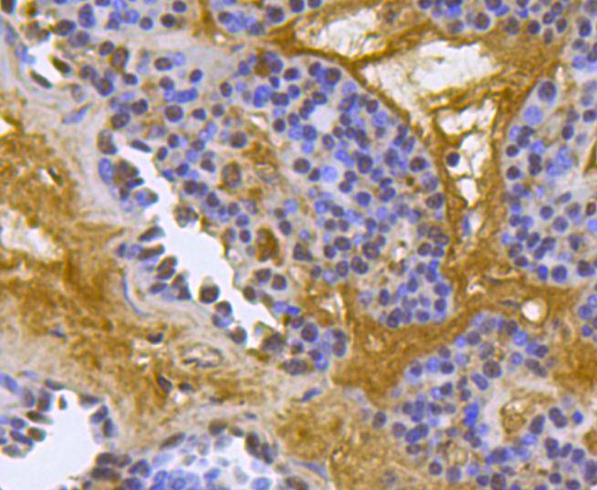
Application
Fig5: Immunohistochemical analysis of paraffin- embedded human liver carcinoma tissue using anti-BMPR1A rabbit polyclonal antibody.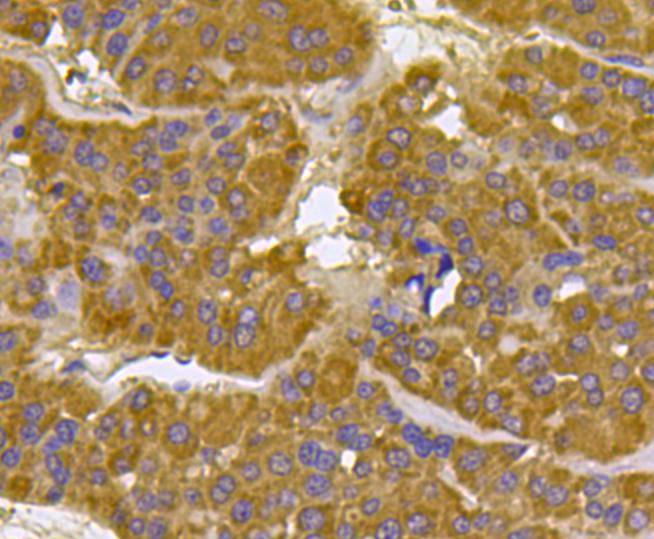
Application
Fig6: Immunohistochemical analysis of paraffin- embedded human liver carcinoma tissue using anti-BMPR1A rabbit polyclonal antibody.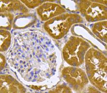
Application
Fig7: Immunohistochemical analysis of paraffin- embedded human kidney tissue using anti-BMPR1A rabbit polyclonal antibody.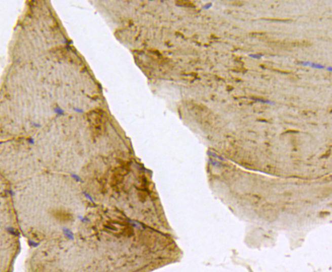
Application
Fig8: Immunohistochemical analysis of paraffin- embedded mouse skeletal muscle tissue using anti-BMPR1A rabbit polyclonal antibody.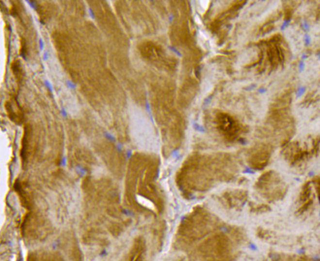
Application
Fig9: Immunohistochemical analysis of paraffin- embedded mouse smooth muscle tissue using anti-BMPR1A rabbit polyclonal antibody.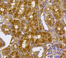
Application
Fig10: Immunohistochemical analysis of paraffin- embedded mouse kidney tissue using anti-BMPR1A rabbit polyclonal antibody.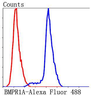
Application
Fig11: Flow cytometric analysis of Jurkat cells with BMPR1A antibody at 1/50 dilution (blue) compared with an unlabelled control (cells without incubation with primary antibody; red). Alexa Fluor 488-conjugated Goat anti mouse IgG was used as the secondary antibody.| Positive Control | Hela, HepG2, SKOV-3, human liver carcinoma tissue, human kidney tissue, mouse skeletal muscle tissue, mouse smooth muscle tissue, mouse kidney tissue. |
|---|---|
| Application Notes | WB:1:1,1000 ICC:1:50-1:200 IHC:1:50-1:200 FC:1:50-1:100 |
| Form | Liquid |
|---|---|
| Storage Instructions | Store at +4℃ after thawing. Aliquot store at -20℃ or -80℃. Avoid repeated freeze / thaw cycles. |
| Storage Buffer | 1*TBS (pH7.4), 1%BSA, 40%Glycerol. Preservative: 0.05% Sodium Azide. |
抱歉,暂无相关文献







