Anti-Histone H3 (tri methyl K4)抗体- ChIP Grade
参阅全部 Histone H3 一抗
兔多克隆抗体to Histone H3 (tri methyl K4) - ChIP Grade
Rabbit
适用于: PepArr, ChIP, WB, IHC-P, ICC/IFmore details
与反应: Cow, Human
预测可用于: Mouse, Rat, Rabbit, Pig, Saccharomyces cerevisiae, Tetrahymena, Xenopus laevis, Arabidopsis thaliana, Caenorhabditis elegans, Drosophila melanogaster, Indian muntjac, Oikopleura, Plants, Zebrafish, Mammals, Trypanosoma cruzi, Common marmoset, Rice, Xenopus tropicalis![]()
Synthetic peptide within Human Histone H3 aa 1-100 (tri methyl K4) conjugated to keyhole limpet haemocyanin. The exact sequence is proprietary.
(Peptide available as ab92374)
ICC/IF: HeLa cells
In immunofluorescence, a distinct property of tri methyl lysine 4 is its apparent 'ringing' of regions that appear as nucleoplasmic 'holes'. These represent the positions of splicing factor compartments, which often are easy to identify using only DNA stains in Indian muntjac fibroblasts. These splicing factor compartments are known to be preferentially associated with active genes and highly acetylated histone H3. This antibody, as expected, fails to stain heterochromatin (work by Kirk McManus, lab of Michael Hendzel).
The immunofluorescence results suggest this antibody is an exceptional euchromatin probe.
Learn about ChIP assay kits, other ChIP antibodies, protocols and more in the ChIP assay guide.
Abcam recommended secondaries - Goat Anti-Rabbit HRP (ab205718) and Goat Anti-Rabbit Alexa Fluor® 488 (ab150077).
The Life Science industry has been in the grips of a reproducibility crisis for a number of years. Abcam is leading the way in addressing this with our range of recombinant monoclonal antibodies and knockout edited cell lines for gold-standard validation. Please check that this product meets your needs before purchasing.
If you have any questions, special requirements or concerns, please send us an inquiry and/or contact our Support team ahead of purchase. Recommended alternatives for this product can be found below, along with publications, customer reviews and Q&As
Liquid
Shipped at 4°C. Store at +4°C short term (1-2 weeks). Upon delivery aliquot. Store at -20°C or -80°C. Avoid freeze / thaw cycle.
pH: 7.40
Preservative: 0.02% Sodium azide
Constituents: 1% BSA, 98.98% PBS
Batches of this product that have a concentration < 1mg/ml may have BSA added as a stabilising agent. If you would like information about the formulation of a specific lot, please contact our scientific support team who will be happy to help.
浓度
批次浓度范围 50 µg 浓度为 0.1 - 1 mg/ml
Immunogen affinity purified
多克隆
IgG
Abpromise™承诺保证使用ab8580于以下的经测试应用
“应用说明”部分 下显示的仅为推荐的起始稀释度;实际最佳的稀释度/浓度应由使用者检定。
| 应用 | Ab评论 | 说明 |
|---|---|---|
| PepArr | Use a concentration of 0.2 - 0.02 µg/ml. Slight cross reactivity is observed with the Histone H3 - di methyl K4 modification. Optimisation is recommended to avoid array signal saturation. | |
| ChIP | (28) | Use 2 µg for 25 µg of chromatin. We recommend GAPDH positive control ChIP primer pair ab267832 as positive control. |
| WB | (20) | Use a concentration of 1 µg/ml. Detects a band of approximately 17 kDa (predicted molecular weight: 15 kDa). |
| IHC-P | (8) | Use at an assay dependent concentration. |
| ICC/IF | (24) | Use a concentration of 1 µg/ml. 1/100 - 1/5000 |
Entrez Gene: 8350 Human
Entrez Gene: 8351 Human
Entrez Gene: 8352 Human
Entrez Gene: 8353 Human
Entrez Gene: 8354 Human
Entrez Gene: 8355 Human
Entrez Gene: 8356 Human
Entrez Gene: 8357 Human
Entrez Gene: 8358 Human
Entrez Gene: 8968 Human
Entrez Gene: 319152 Mouse
Entrez Gene: 319153 Mouse
Entrez Gene: 360198 Mouse
Entrez Gene: 97908 Mouse
Omim: 602810 Human
SwissProt: P68431 Human
SwissProt: P68433 Mouse
Unigene: 132854 Human
Unigene: 247813 Human
Unigene: 247814 Human
Unigene: 248176 Human
Unigene: 443021 Human
Unigene: 484990 Human
Unigene: 532144 Human
Unigene: 533292 Human
Unigene: 546315 Human
Unigene: 586261 Human
Unigene: 591778 Human
Unigene: 221301 Mouse
Unigene: 261657 Mouse
Unigene: 377874 Mouse
Unigene: 390558 Mouse
Unigene: 397328 Mouse
Unigene: 138090 Rat
H3 histone family member E pseudogene antibody
H3 histone family, member A antibody
H3/A antibody
H31_HUMAN antibody
H3F3 antibody
H3FA antibody
Hist1h3a antibody
HIST1H3B antibody
HIST1H3C antibody
HIST1H3D antibody
HIST1H3E antibody
HIST1H3F antibody
HIST1H3G antibody
HIST1H3H antibody
HIST1H3I antibody
HIST1H3J antibody
HIST3H3 antibody
histone 1, H3a antibody
Histone cluster 1, H3a antibody
Histone H3 3 pseudogene antibody
Histone H3.1 antibody
Histone H3/a antibody
Histone H3/b antibody
Histone H3/c antibody
Histone H3/d antibody
Histone H3/f antibody
Histone H3/h antibody
Histone H3/i antibody
Histone H3/j antibody
Histone H3/k antibody
Histone H3/l antibody

ChIP - Anti-Histone H3 (tri methyl K4) antibody - ChIP Grade (ab8580)This image is courtesy of an anonymous Abreview
ChIP was performed with human thyroid cancer cell lysate and 1 µg/µg of ab8580. Lysates were incubated with the primary antibody for 16 hours at 4°C. positive control promoter: GAPDH promoter; positive control enhancer: enhancer region of a published target (10.1093/nar/gkx802); negative control: published negative region (10.1093/nar/gkx802).

Immunocytochemistry/ Immunofluorescence - Anti-Histone H3 (tri methyl K4) antibody - ChIP Grade (ab8580)
ab8580 staining Histone H3 (tri methyl K4) in HeLa cells. The cells were fixed with 100% methanol (5 min), permeabilized with 0.1% PBS-Triton X-100 for 5 minutes and then blocked with 1% BSA/10% normal goat serum/0.3M glycine in 0.1%PBS-Tween for 1h. The cells were then incubated overnight at 4°C with ab8580 at 0.1µg/ml and ab7291, Mouse monoclonal [DM1A] to alpha Tubulin - Loading Control. Cells were then incubated with ab150081, Goat polyclonal Secondary Antibody to Rabbit IgG - H&L (Alexa Fluor® 488), pre-adsorbed at 1/1000 dilution (shown in green) and ab150120, Goat polyclonal Secondary Antibody to Mouse IgG - H&L (Alexa Fluor® 594), pre-adsorbed at 1/1000 dilution (shown in pseudocolour magenta). Nuclear DNA was labelled with DAPI (shown in blue). Also suitable in cells fixed with 4% paraformaldehyde (10 min).Image was acquired with a high-content analyser (Operetta CLS, Perkin Elmer) and a maximum intensity projection of confocal sections is shown.

ChIP - Anti-Histone H3 (tri methyl K4) antibody - ChIP Grade (ab8580)
Chromatin was prepared from U-2 OS (Human bone osteosarcoma epithelial cell line) cells according to the Abcam X-ChIP protocol.
Cells were fixed with formaldehyde for 10 minutes. The ChIP was performed with 25 µg of chromatin, 2 µg of ab8580 (blue), and 20 µl of Protein A/G sepharose beads. No antibody was added to the beads control (yellow). The immunoprecipitated DNA was quantified by real time PCR (Taqman approach). Primers and probes are located in the first kb of the transcribed region.

Western blot - Anti-Histone H3 (tri methyl K4) antibody - ChIP Grade (ab8580)
Anti-Histone H3 (tri methyl K4) antibody - ChIP Grade (ab8580) at 1 µg/ml + Calf thymus histone preparation (nuclear lysate) at 0.5 µg
Secondary
Goat Anti-Rabbit IgG (H+L) HRP- conjugated antibody at 1/50000 dilution
Performed under reducing conditions.
Predicted band size: 15 kDa
Observed band size: 17 kDawhy is the actual band size different from the predicted?
Exposure time: 8 minutes

Immunocytochemistry/ Immunofluorescence - Anti-Histone H3 (tri methyl K4) antibody - ChIP Grade (ab8580)This image is courtesy of Ahmad Khalil and Daniel Driscoll, University of Florida College of Medicine.
Human female lymphoblast immunostained with ab8580 (1:100) (yellowish green) specific for histone H3 lysine 4 (H3-K4) trimethylation; the DNA is stained red with propidium iodide (PI).
Note the inactive X chromosome (arrow) and pericentromeric heterochromatin are largely devoid of this modification.

Immunohistochemistry (Formalin/PFA-fixed paraffin-embedded sections) - Anti-Histone H3 (tri methyl K4) antibody - ChIP Grade (ab8580)
IHC image of ab8580 staining Histone H3 (tri methyl K4) in human colon formalin-fixed paraffin-embedded tissue sections*, performed on a Leica Bond.
The section was pre-treated using heat mediated antigen retrieval with sodium citrate buffer (pH 6, epitope retrieval solution 1) for 20 minutes. The section was then incubated with ab8580, 1/500 dilution, for 15 minutes at room temperature and detected using an HRP conjugated compact polymer system. DAB was used as the chromogen. The section was then counterstained with haematoxylin and mounted with DPX.
No primary antibody was used in the negative control (inset).
For other IHC staining systems (automated and non-automated) customers should optimize variable parameters such as antigen retrieval conditions, primary antibody concentration and antibody incubation times.
*Tissue obtained from the Human Research Tissue Bank, supported by the NIHR Cambridge Biomedical Research Centre
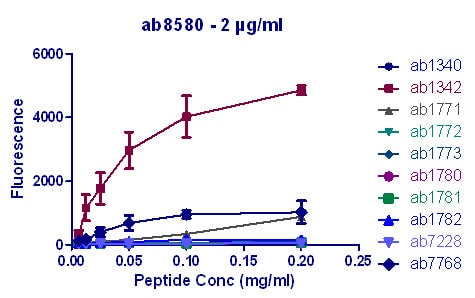
Peptide Array - Anti-Histone H3 (tri methyl K4) antibody - ChIP Grade (ab8580)
All batches of ab8580 are tested in Peptide Array against peptides to different Histone H3 modifications. Six dilutions of each peptide are printed on to the Peptide Array in triplicate and results are averaged before being plotted on to a graph. Results show strong binding to Histone H3 - tri methyl K4 peptide (ab1342), indicating that this antibody specifically recognises the Histone H3 - tri methyl K4 modification. Slight cross reactivity is observed with the Histone H3 - di methyl K4 modification. Optimization is recommended to avoid array signal saturation.
ab1340 - Histone H3 - mono methyl K4
ab1342 - Histone H3 - tri methyl K4
ab1771 - Histone H3 - mono methyl K9
ab1772 - Histone H3 - di methyl K9
ab1773 - Histone H3 - tri methyl K9
ab1780 - Histone H3 - mono methyl K27
ab1781 - Histone H3 - di methyl K27
ab1782 - Histone H3 - tri methyl K27
ab7228 - Histone H3 - unmodified
ab7768 - Histone H3 - di methyl K4

ChIP - Anti-Histone H3 (tri methyl K4) antibody - ChIP Grade (ab8580)This image is courtesy of an anonymous Abreview
Chromatin was prepared from human cell lysate - nuclear B cells according to the Abcam X-ChIP protocol.
Cells were fixed with formaldehyde for 10 minutes. The ChIP was performed with 0.5 µg of ab8580 per µg chromatin in ChIP Buffer fot 16 hours at 4°C. The immunoprecipitated DNA was quantified by real time PCR (Taqman approach). Primers and probes are located in the first kb of the transcribed region. Negative control: IgG and Gene Desert. Positive control: GAPDH Promoter.
抱歉,暂无浏览记录


