Anti-alpha Internexin抗体
参阅全部 alpha Internexin 一抗
兔多克隆抗体to alpha Internexin
Rabbit
Specifically recognizes a-internexin.
适用于: WB, IHC-FoFr, IHC-P, ICC/IFmore details
与反应: Mouse, Rat, Cow, Human
预测可用于: Mammals![]()
Recombinant full length protein corresponding to Rat alpha Internexin.
Database link: P23565
The Life Science industry has been in the grips of a reproducibility crisis for a number of years. Abcam is leading the way in addressing this with our range of recombinant monoclonal antibodies and knockout edited cell lines for gold-standard validation. Please check that this product meets your needs before purchasing.
If you have any questions, special requirements or concerns, please send us an inquiry and/or contact our Support team ahead of purchase. Recommended alternatives for this product can be found below, along with publications, customer reviews and Q&As
Liquid
Shipped at 4°C. Store at +4°C short term (1-2 weeks). Upon delivery aliquot. Store at -20°C or -80°C. Avoid freeze / thaw cycle.
pH: 7.60
Preservatives: 0.01% Thimerosal (merthiolate), 0.05% Sodium azide
Constituents: 0.164% Sodium phosphate, 1.45% Sodium chloride, 1.5% BSA
Whole antiserum
多克隆
IgG
Abpromise™承诺保证使用ab7259于以下的经测试应用
“应用说明”部分 下显示的仅为推荐的起始稀释度;实际最佳的稀释度/浓度应由使用者检定。
| 应用 | Ab评论 | 说明 |
|---|---|---|
| WB | (1) | 1/10000 - 1/20000. Predicted molecular weight: 66 kDa. |
| IHC-FoFr | 1/500 - 1/1000. | |
| IHC-P | 1/2000. | |
| ICC/IF | 1/500 - 1/1000. |
Entrez Gene: 9118 Human
Entrez Gene: 226180 Mouse
Omim: 605338 Human
SwissProt: Q16352 Human
SwissProt: P46660 Mouse
Unigene: 500916 Human
Unigene: 276251 Mouse
Unigene: 88941 Rat
66 kDa neurofilament protein antibody
AINX_HUMAN antibody
Alpha Inx antibody
Alpha-internexin antibody
Alpha-Inx antibody
INA antibody
Internexin neuronal intermediate filament protein alpha antibody
MGC12702 antibody
NEF 5 antibody
NEF5 antibody
Neurofilament 5 (66kD) antibody
Neurofilament 5 antibody
Neurofilament 66 antibody
Neurofilament 66 tax binding protein antibody
Neurofilament-66 antibody
NF 66 antibody
NF-66 antibody
NF66 antibody
TXBP 1 antibody
TXBP1 antibody

Western blot - Anti-alpha Internexin antibody (ab7259)
All lanes : Anti-alpha Internexin antibody (ab7259) at 1/10000 dilution
Lane 1 : Protein standard
Lane 2 : Mouse spinal cord lysate
Lane 3 : Rat spinal cord lysate
Lane 4 : Cow spinal cord lysate
Predicted band size: 66 kDa

Immunohistochemistry (PFA perfusion fixed frozen sections) - Anti-alpha Internexin antibody (ab7259)
Immunofluorescence analysis of rat cerebellum section stained for alpha Internexin using ab7259 at a 1/2000 dilution (green) co-strained with chicken polyclonal antibody to GFAP at a 1/5000 dilution (red). DAPI was used to stain nuclear DNA (blue).
Following transcardial perfusion with 4% paraformaldehyde, brain was post-fixed for 24 hours, cut to 45 μM, and free-floating sections were stained.

Immunocytochemistry/ Immunofluorescence - Anti-alpha Internexin antibody (ab7259)
Ab7259 shows red, neuronal progenitor cells, plectin (not one of our antibodies) shows fibroblast marker. Photo courtesy of: Dr. Gerry Shaw University of Florida
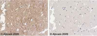
Immunohistochemistry (Formalin/PFA-fixed paraffin-embedded sections) - Anti-alpha Internexin antibody (ab7259)
Ab7259 staining Human normal temporal cortex. Staining is localised to intracellualr compartment.
Left panel: with primary antibody diluted 1:2000. Right panel: isotype control.
Sections were stained using an automated system DAKO Autostainer Plus , at room temperature: sections were rehydrated and antigen retrieved with the Dako 3 in 1 AR buffer citrate pH6.1 in a DAKO PT Link. Slides were peroxidase blocked in 3% H2O2 in methanol for 10 mins. They were then blocked with Dako Protein block for 10 minutes (containing casein 0.25% in PBS) then incubated with primary antibody for 20 min and detected with Dako envision flex amplification kit for 30 minutes. Colorimetric detection was completed with Diaminobenzidine for 5 minutes. Slides were counterstained with Haematoxylin and coverslipped under DePeX. Please note that for manual staining we recommend to optimize the primary antibody concentration and incubation time (overnight incubation), and amplification may be required.

Immunocytochemistry/ Immunofluorescence - Anti-alpha Internexin antibody (ab7259)
ICC/IF image of ab7259 stained PC12 cells. The cells were 100% methanol fixed (5 min) and then incubated in 1%BSA / 10% normal goat serum / 0.3M glycine in 0.1% PBS-Tween for 1h to permeabilise the cells and block non-specific protein-protein interactions. The cells were then incubated with the antibody (ab7259, 1/1000 dilution) overnight at +4°C. The secondary antibody (green) was Alexa Fluor® 488 goat anti-rabbit IgG (H+L) used at a 1/1000 dilution for 1h. Alexa Fluor® 594 WGA was used to label plasma membranes (red) at a 1/200 dilution for 1h. DAPI was used to stain the cell nuclei (blue) at a concentration of 1.43µM.
抱歉,暂无浏览记录


