生物素Anti-Collagen I抗体
参阅全部 Collagen I 一抗
生物素兔多克隆抗体to Collagen I
Rabbit
Biotin
Some class-specific anti-collagens may be specific for three-dimensional epitopes which may result in diminished reactivity with denatured collagen or formalin-fixed, paraffin embedded tissues.  This antibody reacts with most mammalian Type I collagens and has expected cross-reactivity with Type III and negligible cross reactivity with Type II, IV, V or VI collagens. Non-specific cross-reaction of anti-collagen antibodies with other human serum proteins or non-collagen extracellular matrix proteins has not been tested.
适用于: IHC-P, Flow Cyt (Intra)more details
与反应: Human
预测可用于: Mammals![]()
Full length native protein (purified) corresponding to Collagen I. Collagen Type I from human and bovine placenta.
Flow Cyt (Intra): Primary adult human dermal fibroblast cells.
At least 11 genetically distinct gene products are collectively referred to as 'collagen types' or other proteins and proteoglycans of the extracellular matrix. In humans, collagens are composed of about 20 unique protein chains which under go various types of post-translational modifications and are ultimately assembled into a triple helix. This results in great diversity between collagen types. Collagens are highly conserved throughout evolution and are characterized by an uninterrupted "Glycine-X-Y" triplet repeat that is a necessary part of the triple helical structure. For these reasons it is often extremely difficult to generate antibodies with specificities to collagens. The development of type specific antibodies is dependent on NON-DENATURED three-dimensional epitopes. This preparation results in a native conformation of the protein.
This antibody is well suited to detect extracellular matrix proteins in normal as well as disease state tissues. Disruption of tissue organization is the hallmark of neoplasia. Malignant lesions can be distinguished from benign by examining the breakdown of basement membranes and loss of 3-dimensional architecture. Malignant cells are presumed to use matrix metalloproteases to degrade barriers created by the extracellular matrix which then allows metastasis to occur. Collagenases, stomelysins and gelatinases can collectively degrade all of the various components of the extracellular matrix, including fibrillar and non-fibrillar collagens and basement membrane glycoproteins.
The Life Science industry has been in the grips of a reproducibility crisis for a number of years. Abcam is leading the way in addressing this with our range of recombinant monoclonal antibodies and knockout edited cell lines for gold-standard validation. Please check that this product meets your needs before purchasing.
If you have any questions, special requirements or concerns, please send us an inquiry and/or contact our Support team ahead of purchase. Recommended alternatives for this product can be found below, along with publications, customer reviews and Q&As
Liquid
Shipped at 4°C. Store at +4°C short term (1-2 weeks). Store at -20°C or -80°C. Avoid freeze / thaw cycle.
Preservative: 0.01% Sodium azide
Constituents: 1% BSA, 0.424% Potassium phosphate solution, 0.88% Sodium chloride
浓度
100 µg 浓度为 1 mg/ml
Immunogen affinity purified
This product has been prepared by immunoaffinity chromatography using immobilized antigens.
This antibody is well suited to detect extracellular matrix proteins in normal as well as disease state tissues. Disruption of tissue organization is the hallmark of neoplasia. Malignant lesions can be distinguished from benign by examining the breakdown of basement membranes and loss of 3-dimensional architecture. Malignant cells are presumed to use matrix metalloproteases to degrade barriers created by the extracellular matrix which then allows metastasis to occur. Collagenases, stomelysins and gelatinases can collectively degrade all of the various components of the extracellular matrix, including fibrillar and non-fibrillar collagens and basement membrane glycoproteins.
多克隆
IgG
Abpromise™承诺保证使用ab6577于以下的经测试应用
“应用说明”部分 下显示的仅为推荐的起始稀释度;实际最佳的稀释度/浓度应由使用者检定。
| 应用 | Ab评论 | 说明 |
|---|---|---|
| IHC-P | 1/50 - 1/200. | |
| Flow Cyt (Intra) | Use at an assay dependent concentration. |
Entrez Gene: 1277 Human
Entrez Gene: 1278 Human
Omim: 120150 Human
Omim: 120160 Human
SwissProt: P02452 Human
SwissProt: P08123 Human
Unigene: 172928 Human
Unigene: 681002 Human
alpha 2(I) procollagen antibody
alpha 2(I)-collagen antibody
Alpha-1 type I collagen antibody
alpha1(I) procollagen antibody
CO1A1_HUMAN antibody
COL1A1 antibody
COL1A2 antibody
collagen 1 antibody
collagen alpha 1 chain type I antibody
Collagen alpha-1(I) chain antibody
collagen alpha-1(I) chain preproprotein antibody
Collagen I alpha 1 polypeptide antibody
Collagen I alpha 2 polypeptide antibody
collagen of skin, tendon and bone, alpha-1 chain antibody
collagen of skin, tendon and bone, alpha-2 chain antibody
Collagen type I alpha 1 antibody
Collagen type I alpha 2 antibody
EDSC antibody
OI1 antibody
OI2 antibody
OI3 antibody
OI4 antibody
pro-alpha-1 collagen type 1 antibody
type I proalpha 1 antibody
type I procollagen alpha 1 chain antibody
Type I procollagen antibody
Alpha 1 type I collagen antibody
Alpha 2 type I collagen antibody
alpha 2 type I procollagen antibody

Immunohistochemistry (Formalin/PFA-fixed paraffin-embedded sections) - Biotin Anti-Collagen I antibody (ab6577)
Immunohistochemistry (Formalin/PFA-fixed paraffin-embedded sections) analysis of human skin tissue sections at pH9 labeling Collagen I with ab6577 10 µg/mL for 1 h at RT. Secondary antibody: Peroxidase rabbit secondary antibody at 1/10,000 for 45 min at RT. Localization: Collagen Type I is secreted in the extracellular matrix. Staining: Collagen Type I as precipitated brown signal (A) with hematoxylin purple nuclear counterstain. With corresponding negative conrol (B).
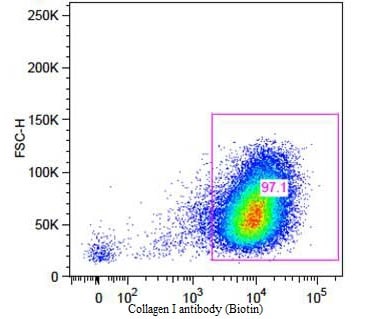
Flow Cytometry (Intracellular) - Biotin Anti-Collagen I antibody (ab6577)
Flow Cytometry analysis of primary adult human dermal fibroblast cells labeling Collagen I with ab6577 5µg/mL for 45 min at 4°C. Secondary antibody: Rabbit Streptavidin, R-PE antibody at 1/500 for 15 min at RT.

Immunohistochemistry (Formalin/PFA-fixed paraffin-embedded sections) - Biotin Anti-Collagen I antibody (ab6577)
Immunohistochemistry (Formalin/PFA-fixed paraffin-embedded sections) analysis of human skin tissue sections at pH6 labeling Collagen I with ab6577 10 µg/mL for 1 h at RT. Secondary antibody: Peroxidase rabbit secondary antibody at 1/10,000 for 45 min at RT. Localization: Collagen Type I is secreted in the extracellular matrix. Staining: Collagen Type I as precipitated brown signal (A) with hematoxylin purple nuclear counterstain. With corresponding negative conrol (B).
抱歉,暂无浏览记录


