Anti-IGF1 Receptor (phospho Y1158 + Y1162 + Y1163)抗体
兔多克隆抗体to IGF1 Receptor (phospho Y1131 + Y1135 + Y1136) + Insulin Receptor (phospho Y1158 + Y1162 + Y1163)
Rabbit
适用于: WBmore details
与反应: Human
预测可用于: Mouse, Rat![]()
Synthetic peptide corresponding to IGF1 Receptor (phospho Y1158 + Y1162 + W1163).
WB: CHO-T (Chinese hamster ovary cell line) whole cell extract transfected with a vector encoding the human insulin receptor and stimulated with insulin.
The antiserum was produced against a chemically synthesized phosphopeptide derived from the region of IR/IGF1R that contains tyrosines 1158, 1162 and 1163 of the human insulin receptor (IR) as numbered according to Ebina, et al. (1146, 1150 and 1151 according to Ullrich, et al.). The corresponding residues in the IGF1R are 1131, 1135 and 1136. The sequence is conserved in human, mouse and rat for both the IR and IGF1R.The two relevant papers are:Ebina, Y., et al. (1985) The human insulin receptor cDNA: the structural basis for hormone-activated transmembrane signalling. Cell 40(4):747-758.Ullrich, A., et al. (1985) Human insulin receptor and its relationship to the tyrosine kinase family of oncogenes. Nature 313(6005):756-761. There are a long and a short isoform of this protein. This is why we are listing 1158, 1162 and 1163 in the name (where these phospho sites in the long isoform) as well as 1146, 1150 and 1151 (for the short isoform).
The Life Science industry has been in the grips of a reproducibility crisis for a number of years. Abcam is leading the way in addressing this with our range of recombinant monoclonal antibodies and knockout edited cell lines for gold-standard validation. Please check that this product meets your needs before purchasing.
If you have any questions, special requirements or concerns, please send us an inquiry and/or contact our Support team ahead of purchase. Recommended alternatives for this product can be found below, along with publications, customer reviews and Q&As
Liquid
Shipped at 4°C. Upon delivery aliquot and store at -20°C or -80°C. Avoid repeated freeze / thaw cycles.
pH: 7.30
Preservative: 0.05% Sodium azide
Constituents: PBS, 50% Glycerol (glycerin, glycerine), 0.1% BSA
浓度
50 µl 浓度为 0.8 mg/ml
Immunogen affinity purified
The antibody has been negatively preadsorbed using a non-phosphopeptide corresponding to the site of phosphorylation to remove antibody that is reactive with non-phosphorylated Insulin Receptor (IR). The final product is generated by affinity chromatography using an IR-derived peptide phosphorylated at tyrosines 1158, 1162 and 1163 (1131, 1135 and 1136 for IGF1R).
多克隆
IgG
Abpromise™承诺保证使用ab5681于以下的经测试应用
“应用说明”部分 下显示的仅为推荐的起始稀释度;实际最佳的稀释度/浓度应由使用者检定。
| 应用 | Ab评论 | 说明 |
|---|---|---|
| WB | (1) | 1/1000. Detects a band of approximately 100 kDa. |
Entrez Gene: 3480 Human
Entrez Gene: 3643 Human
Entrez Gene: 16001 Mouse
Entrez Gene: 16337 Mouse
Omim: 147370 Human
Omim: 147670 Human
SwissProt: P06213 Human
SwissProt: P08069 Human
SwissProt: P15208 Mouse
SwissProt: Q60751 Mouse
Unigene: 465744 Human
Unigene: 643120 Human
Unigene: 714012 Human
Unigene: 268003 Mouse
Unigene: 275742 Mouse
Unigene: 10957 Rat
Unigene: 165078 Rat
Unigene: 9876 Rat
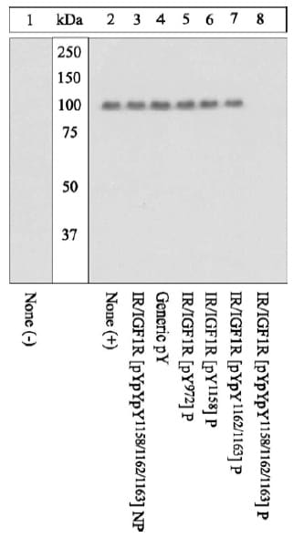
Western blot - Anti-IGF1 Receptor (phospho Y1158 + Y1162 + Y1163) antibody (ab5681)
All lanes : Anti-IGF1 Receptor (phospho Y1158 + Y1162 + Y1163) antibody (ab5681) at 1/1000 dilution (2 hours at room temperature in a 3% BSA-TBST buffer)
Lane 1 : Unstimulated (-), CHO-T transfected with insulin receptor containing vector whole cell extract with 5% BSA TBST buffer overnight at 4°C
Lanes 2-8 : Insulin stimulated (+), CHO-T transfected with insulin receptor containing vector whole cell extract with 5% BSA TBST buffer overnight at 4°C
Secondary
All lanes : goat F(ab’ 2 anti-rabbit IgG HRP conjugate
Peptide Competition - prior primary antibody incubation:
1 and 2 - no peptide,
3 - non-phosphorylated peptide corresponding to the immunogen,
4 - generic phosphotyrosine-containing peptide,
5 to 7 - phosphopeptides corresponding to other IR/IGF1R sites,
8 - phosphopeptide immunogen.
10% SDS-PAGE transferred to PVDF.
Method of detection: Pierce SuperSignal method.
The data show that only the phosphopeptide corresponding to IR/IGF1R [pYpYpY1158/1162/1163] completely blocks the antibody signal, thereby demonstrating the specificty.


