Anti-EGFR (phospho Y1173)抗体
参阅全部 EGFR 一抗
兔多克隆抗体to EGFR (phospho Y1173)
Rabbit
适用于: WB, ICC, IHC-Pmore details
与反应: Mouse, Human
Synthetic peptide corresponding to Human EGFR (phospho Y1173).
WB: NIH/3T3 cells. IHC-P: Human hepatocellular carcinoma, human lung adenocarcinoma. ICC: HeLa cells.
The Life Science industry has been in the grips of a reproducibility crisis for a number of years. Abcam is leading the way in addressing this with our range of recombinant monoclonal antibodies and knockout edited cell lines for gold-standard validation. Please check that this product meets your needs before purchasing.
If you have any questions, special requirements or concerns, please send us an inquiry and/or contact our Support team ahead of purchase. Recommended alternatives for this product can be found below, along with publications, customer reviews and Q&As
Liquid
Shipped at 4°C. Upon delivery aliquot and store at -20°C. Avoid freeze / thaw cycles.
pH: 7.30
Preservative: 0.05% Sodium azide
Constituents: PBS, 50% Glycerol, 0.1% BSA
浓度
50 µl 浓度为 0.09 mg/ml
Immunogen affinity purified
The antibody has been negatively preadsorbed using (i) a non phosphopeptide corresponding to the site of phosphorylation to remove antibody that is reactive with non-phosphorylated epidermal growth factor receptor (EGFR), and (ii) a generic tyrosine phosphorylated peptide to remove antibody that is reactive with phosphotyrosine, irrespective of the sequence. The final product is generated by affinity chromatography using an EGFR-derived peptide that is phosphorylated at tyrosine 1173.
多克隆
IgG
Abpromise™承诺保证使用ab5652于以下的经测试应用
“应用说明”部分 下显示的仅为推荐的起始稀释度;实际最佳的稀释度/浓度应由使用者检定。
| 应用 | Ab评论 | 说明 |
|---|---|---|
| WB | (1) | 1/1000. Detects a band of approximately 185 kDa. |
| ICC | 1/100 - 1/500. | |
| IHC-P | 1/10 - 1/100. |
Entrez Gene: 1956 Human
Entrez Gene: 13649 Mouse
Omim: 131550 Human
SwissProt: P00533 Human
SwissProt: Q01279 Mouse
Unigene: 488293 Human
Unigene: 605083 Human
Unigene: 420648 Mouse
Unigene: 439882 Mouse
Unigene: 8534 Mouse
Avian erythroblastic leukemia viral (v erb b) oncogene homolog antibody
Cell growth inhibiting protein 40 antibody
Cell proliferation inducing protein 61 antibody
EGF R antibody
EGFR antibody
EGFR_HUMAN antibody
Epidermal growth factor receptor (avian erythroblastic leukemia viral (v erb b) oncogene homolog) antibody
Epidermal growth factor receptor (erythroblastic leukemia viral (v erb b) oncogene homolog avian) antibody
Epidermal growth factor receptor antibody
erb-b2 receptor tyrosine kinase 1 antibody
ERBB antibody
ERBB1 antibody
Errp antibody
HER1 antibody
mENA antibody
NISBD2 antibody
Oncogen ERBB antibody
PIG61 antibody
Proto-oncogene c-ErbB-1 antibody
Receptor tyrosine protein kinase ErbB 1 antibody
Receptor tyrosine-protein kinase ErbB-1 antibody
SA7 antibody
Species antigen 7 antibody
Urogastrone antibody
v-erb-b Avian erythroblastic leukemia viral oncogen homolog antibody
wa2 antibody
Wa5 antibody

Immunocytochemistry - Anti-EGFR (phospho Y1173) antibody (ab5652)
HeLa cells stained for EGFR (green) using ab5652 at 2 µg/mL in ICC/IF. Followed by Alexa Fluor 488 Goat Anti-Rabbit IgG Secondary Antibody at 1/400 dilution for 30 minutes at room temperature (Panel a). Nuclei (Panel b: blue) were stained with SlowFade® Gold Antifade Mountant with DAPI. F-actin (Panel c: red) was stained with Alexa Fluor 594 Phalloidin. Panel d is a merged image showing cytoplasmic localization of EGFR (pY1173). Panel e shows untreated cells. Panel f shows no primary antibody control.

Immunohistochemistry (Formalin/PFA-fixed paraffin-embedded sections) - Anti-EGFR (phospho Y1173) antibody (ab5652)
Paraffin embedded human lung adenocarcinoma tissue stained for EGFR using ab5652 at 1/50 dilution in immunohistochemical analysis.

Immunohistochemistry (Formalin/PFA-fixed paraffin-embedded sections) - Anti-EGFR (phospho Y1173) antibody (ab5652)
Paraffin embedded human hepatocellular carcinoma tissue stained for EGFR using ab5652 at 1/20 dilution in immunohistochemical analysis.
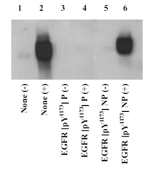
Western blot - Anti-EGFR (phospho Y1173) antibody (ab5652)
Cell extracts prepared from NIH3T3 cells expressing EGFR were starved for 30 hours, then stimulated for 10 minutes with 30 ng/mL EGF (+), or left unstimulated (-), then resolved by SDS-PAGE on a 6% Tris-glycine gel, and transferred to nitrocellulose. Membranes were incubated with 0.50 µg/mL ab5652 antibody, following prior incubation in the absence (lanes 1& 2), or presence of the peptide immunogen (lanes 3 & 4), or the nonphosphopeptide corresponding to the EGFR phosphopeptide (lanes 5 & 6). After washing, membranes were incubated with goat F(ab’)2 anti-rabbit IgG alkaline phosphatase and bands were detected using the Tropix WesternStar detection method. The data show that only the phosphopeptide corresponding to this site blocks the antibody signal, demonstrating the specificity of the ab5652 antibody for this phosphorylated residue. Cell extracts prepared from NIH3T3 cells expressing EGFR were starved for 30 hours, then stimulate


