Anti-EGFR (phospho Y992)抗体
参阅全部 EGFR 一抗
兔多克隆抗体to EGFR (phospho Y992)
Rabbit
适用于: WB, IHC-P, ICCmore details
与反应: Human
Synthetic peptide corresponding to Human EGFR (phospho Y992).
A431 cells stimulated with EGF; human lung adenocarcinoma, human hepatocellular carcinoma
The Life Science industry has been in the grips of a reproducibility crisis for a number of years. Abcam is leading the way in addressing this with our range of recombinant monoclonal antibodies and knockout edited cell lines for gold-standard validation. Please check that this product meets your needs before purchasing.
If you have any questions, special requirements or concerns, please send us an inquiry and/or contact our Support team ahead of purchase. Recommended alternatives for this product can be found below, along with publications, customer reviews and Q&As
Liquid
Shipped at 4°C. Upon delivery aliquot and store at -20°C. Avoid freeze / thaw cycles.
pH: 7.30
Preservative: 0.05% Sodium azide
Constituents: PBS, 50% Glycerol (glycerin, glycerine), 0.1% BSA
PBS is without Mg2+ and Ca2+ and BSA is IgG and protease free.
浓度
50 µl 浓度为 0.5 mg/ml
Immunogen affinity purified
The antibody has been negatively preadsorbed using (i) a non phosphopeptide corresponding to the site of phosphorylation to remove antibody that is reactive with non-phosphorylated epidermal growth factor receptor (EGFR), and (ii) a generic tyrosine phosphorylated peptide to remove antibody that is reactive with phosphotyrosine, irrespective of the sequence. The final product is generated by affinity chromatography using an EGFR-derived peptide that is phosphorylated at tyrosine 922.
多克隆
IgG
Abpromise™承诺保证使用ab5638于以下的经测试应用
“应用说明”部分 下显示的仅为推荐的起始稀释度;实际最佳的稀释度/浓度应由使用者检定。
| 应用 | Ab评论 | 说明 |
|---|---|---|
| WB | 1/1000. Detects a band of approximately 170 kDa. | |
| IHC-P | 1/10 - 1/1000. | |
| ICC | 1/100 - 1/500. |
Entrez Gene: 1956 Human
Omim: 131550 Human
SwissProt: P00533 Human
Unigene: 488293 Human
Unigene: 605083 Human
EGF R antibody
EGFR antibody
EGFR_HUMAN antibody
Epidermal growth factor receptor (avian erythroblastic leukemia viral (v erb b) oncogene homolog) antibody
Epidermal growth factor receptor (erythroblastic leukemia viral (v erb b) oncogene homolog avian) antibody
Epidermal growth factor receptor antibody
erb-b2 receptor tyrosine kinase 1 antibody
ERBB antibody
ERBB1 antibody
Errp antibody
HER1 antibody
mENA antibody
NISBD2 antibody
Oncogen ERBB antibody
PIG61 antibody
Proto-oncogene c-ErbB-1 antibody
Receptor tyrosine protein kinase ErbB 1 antibody
Receptor tyrosine-protein kinase ErbB-1 antibody
SA7 antibody
Species antigen 7 antibody
Urogastrone antibody
v-erb-b Avian erythroblastic leukemia viral oncogen homolog antibody
wa2 antibody
Wa5 antibody
Avian erythroblastic leukemia viral (v erb b) oncogene homolog antibody
Cell growth inhibiting protein 40 antibody
Cell proliferation inducing protein 61 antibody

Western blot - Anti-EGFR (phospho Y992) antibody (ab5638)
All lanes : Anti-EGFR (phospho Y992) antibody (ab5638) at 1/1000 dilution
Lane 1 : A431 cells unstimulated
Lanes 2-5 : A431 cells stimulated with 200 ng/mL EGF for 15 minutes

Immunohistochemistry (Formalin/PFA-fixed paraffin-embedded sections) - Anti-EGFR (phospho Y992) antibody (ab5638)
Immunohistochemistry analysis of EGFR (pY992) with ab5638 at 1/20 dilution showing staining in the cytoplasm and membrane of paraffin-embedded human lung adenocarcinoma (right) compared to a negative control without primary antibody (left).
Antigen retrieval was performed using 10mM sodium citrate (pH 6.0), microwaved for 8-15 min.

Immunohistochemistry (Formalin/PFA-fixed paraffin-embedded sections) - Anti-EGFR (phospho Y992) antibody (ab5638)
Immunohistochemistry analysis of EGFR (pY992) with ab5638 at 1/20 dilution showing staining in the cytoplasm and membrane of paraffin-embedded human hepatocellular carcinoma (right) compared to a negative control without primary antibody (left).
Antigen retrieval was performed using 10mM sodium citrate (pH 6.0), microwaved for 8-15 min.
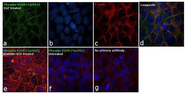
Immunocytochemistry - Anti-EGFR (phospho Y992) antibody (ab5638)
Immunocytochemical analysis of EGFR [pY992] was performed using A-431 cells treated with 200 ng/mL of EGF for 10 minutes. The cells were fixed with 4% paraformaldehyde, permeabilized with 0.1% Triton™ X-100, and blocked with 1% BSA. The cells were labeled with ab5638 at 1/250 dilution and incubated overnight at 4°C followed by a Goat anti-Mouse IgG (H+L) Secondary Antibody, Alexa Fluor® 488 conjugate at a dilution of 1/2000 for 45 minutes at room temperature (Panel a: green). Nuclei (Panel b: blue) DAPI (Panel c: red) F-actin was stained with Rhodamine Phalloidin at 1/300. Panel d represents the merged image showing membranous localization. Panel e represents cells treated with antagonist, Afatinib (1µM for 6hrs) followed by EGF (200 ng/mL for 10 minutes), showing reduced Phospho-EGFR staining. Panel f shows untreated cells with no signal. Panel g represents control cells with no primary antibody to assess background.


