Anti-Hsp27抗体
参阅全部 Hsp27 一抗
兔多克隆抗体to Hsp27
Rabbit
适用于: WB, ICC/IF, IHC-Pmore details
与反应: Mouse, Rat, Human, African green monkey
Synthetic peptide corresponding to Human Hsp27 aa 10-21.
Sequence:
LLRGPSWDPFRC
(Peptide available as ab39789)
WB: HEK-293T, HeLa, K562, A431, HepG2, COS-7, NIH/3T3, MCF7, MDA-MB-231, PC3, DU 145, LNCaP, HT1080 whole cell lysate. IHC-P: Human skeletal muscle and breast carcinoma tissue. ICC/IF: HeLa, MCF-7 and C6 cells.
The Life Science industry has been in the grips of a reproducibility crisis for a number of years. Abcam is leading the way in addressing this with our range of recombinant monoclonal antibodies and knockout edited cell lines for gold-standard validation. Please check that this product meets your needs before purchasing.
If you have any questions, special requirements or concerns, please send us an inquiry and/or contact our Support team ahead of purchase. Recommended alternatives for this product can be found below, along with publications, customer reviews and Q&As
Liquid
Shipped at 4°C. Store at +4°C short term (1-2 weeks). Upon delivery aliquot. Store at -20°C long term. Avoid freeze / thaw cycle.
Preservative: 0.05% Sodium azide
Constituents: 99% PBS, 0.1% BSA
Immunogen affinity purified
Antigen affinity chromatography.
多克隆
IgG
Abpromise™承诺保证使用ab5579于以下的经测试应用
“应用说明”部分 下显示的仅为推荐的起始稀释度;实际最佳的稀释度/浓度应由使用者检定。
| 应用 | Ab评论 | 说明 |
|---|---|---|
| WB | (1) | 1/1000 - 1/2000. Detects a band of approximately 27 kDa. |
| ICC/IF | 1/50. | |
| IHC-P | Use a concentration of 4 µg/ml. Perform heat mediated antigen retrieval via the microwave method before commencing with IHC staining protocol. |
Entrez Gene: 3315 Human
Entrez Gene: 15507 Mouse
Omim: 602195 Human
SwissProt: P04792 Human
SwissProt: P14602 Mouse
Unigene: 520973 Human
Unigene: 13849 Mouse
Unigene: 3841 Rat
Heat shock 27kDa protein antibody
28 kDa heat shock protein antibody
CMT2F antibody
DKFZp586P1322 antibody
epididymis secretory protein Li 102 antibody
Estrogen regulated 24 kDa protein antibody
Estrogen-regulated 24 kDa protein antibody
Heat shock 25kDa protein 1 antibody
Heat shock 27 kDa protein antibody
Heat shock 27kD protein 1 antibody
Heat shock 27kDa protein 1 antibody
Heat shock 28kDa protein 1 antibody
Heat Shock Protein 27 antibody
Heat shock protein beta 1 antibody
Heat shock protein beta-1 antibody
heat shock protein family B (small) member 1 antibody
HEL-S-102 antibody
HMN2B antibody
HS.76067 antibody
Hsp 25 antibody
HSP 27 antibody
Hsp 28 antibody
Hsp B1 antibody
Hsp25 antibody
HSP27 antibody
Hsp28 antibody
HspB1 antibody
HSPB1_HUMAN antibody
SRP27 antibody
Stress responsive protein 27 antibody
Stress-responsive protein 27 antibody

Western blot - Anti-Hsp27 antibody (ab5579)
All lanes : Anti-Hsp27 antibody (ab5579) at 1/2000 dilution
Lane 1 : MCF7 (human breast adenocarcinoma cell line) whole cell lysate
Lane 2 : MDA-MB-231 (human breast adenocarcinoma cell line) whole cell lysate
Lane 3 : PC3 (human prostate adenocarcinoma cell line) whole cell lysate
Lane 4 : DU 145 (human prostate carcinoma cell line) whole cell lysate
Lane 5 : HeLa (human epithelial cell line from cervix adenocarcinoma) whole cell lysate
Lane 6 : LNCaP (human prostate cancer cell line) whole cell lysate
Lane 7 : HT1080 (human fibrosarcoma cell line) whole cell lysate
Lysates/proteins at 30 µg per lane.
Secondary
All lanes : Goat anti-Rabbit IgG (H+L) Superclonal™ Recombinant Secondary Antibody, HRP at 1/4000 dilution
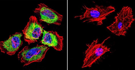
Immunocytochemistry/ Immunofluorescence - Anti-Hsp27 antibody (ab5579)
Immunofluorescence analysis of HeLa (Human epithelial cell line from cervix adenocarcinoma) cells labeling Hsp27 (green) with ab5579 at 1/200 dilution, followed by DyLight 488-conjugated secondary antibody. F-Actin staining with Phalloidin (red) and nuclei with DAPI (blue). Cells were fixed with formaldehyde and incubated with the primary antibody overnight at 4°C. 60X magnification. Right - negative control.

Immunohistochemistry (Formalin/PFA-fixed paraffin-embedded sections) - Anti-Hsp27 antibody (ab5579)
Immunohistochemical analysis of both normal and cancer biopsies of deparaffinized human skeletal muscle tissue labeling Hsp27 with ab5579 at 1/20 dilution or without primary antibody (negative control) overnight at 4°C in a humidified chamber. Tissues were washed extensively with PBST and endogenous peroxidase activity was quenched with a peroxidase suppressor. Detection was performed using a biotin-conjugated secondary antibody and SA-HRP, followed by colorimetric detection using DAB. Tissues were counterstained with hematoxylin and prepped for mounting.
To expose target proteins, heat induced antigen retrieval was performed using 10mM sodium citrate (pH 6.0) buffer, microwaved for 8-15 minutes. Following antigen retrieval tissues were blocked in 3% BSA-PBS for 30 minutes at room temperature.

Immunocytochemistry/ Immunofluorescence - Anti-Hsp27 antibody (ab5579)
Immunofluorescence analysis of MCF7 (Human breast adenocarcinoma cell line) cells labeling Hsp27 (green) with ab5579 at 1/200 dilution, followed by DyLight 488-conjugated secondary antibody. F-Actin staining with Phalloidin (red) and nuclei with DAPI (blue). Cells were fixed with formaldehyde and incubated with the primary antibody overnight at 4°C. 60X magnification. Right - negative control.

Immunohistochemistry (Formalin/PFA-fixed paraffin-embedded sections) - Anti-Hsp27 antibody (ab5579)
Immunohistochemical analysis of both normal and cancer biopsies of deparaffinized human breast carcinoma tissue labeling Hsp27 with ab5579 at 1/100 dilution or without primary antibody (negative control) overnight at 4°C in a humidified chamber. Tissues were washed extensively with PBST and endogenous peroxidase activity was quenched with a peroxidase suppressor. Detection was performed using a biotin-conjugated secondary antibody and SA-HRP, followed by colorimetric detection using DAB. Tissues were counterstained with hematoxylin and prepped for mounting.
To expose target proteins, heat induced antigen retrieval was performed using 10mM sodium citrate (pH6.0) buffer, microwaved for 8-15 minutes. Following antigen retrieval tissues were blocked in 3% BSA-PBS for 30 minutes at room temperature.
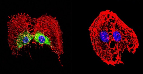
Immunocytochemistry/ Immunofluorescence - Anti-Hsp27 antibody (ab5579)
Immunofluorescence analysis of C6 (Rat glial tumor cell line) cells labeling Hsp27 (green) with ab5579 at 1/100 dilution, followed by DyLight 488-conjugated secondary antibody. F-Actin staining with Phalloidin (red) and nuclei with DAPI (blue). Cells were fixed with formaldehyde and incubated with the primary antibody overnight at 4°C. 60X magnification. Right - negative control.

Western blot - Anti-Hsp27 antibody (ab5579)
All lanes : Anti-Hsp27 antibody (ab5579) at 1/1000 dilution
Lane 1 : HEK-293T (human epithelial cell line from embryonic kidney transformed with large T antigen) whole cell lysate
Lane 2 : HeLa (human epithelial cell line from cervix adenocarcinoma) whole cell lysate
Lane 3 : K562 (human chronic myelogenous leukemia lymphoblast cell line ) whole cell lysate
Lane 4 : A431 (human epidermoid carcinoma cell line) whole cell lysate
Lane 5 : HepG2 (human liver hepatocellular carcinoma cell line) whole cell lysate
Lane 6 : COS-7 (african green monkey kidney fibroblast-like cell line) whole cell lysate
Lane 7 : NIH/3T3 (mouse embryonic fibroblast cell line) whole cell lysate
Lysates/proteins at 50 µg per lane.
Secondary
All lanes : Goat anti-rabbit IgG-HRP secondary antibody at 1/20000 dilution
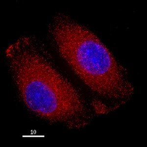
Immunocytochemistry/ Immunofluorescence - Anti-Hsp27 antibody (ab5579)This image is courtesy of Michael Manicini, Ph.D.
HeLa (human epithelial cell line from cervix adenocarcinoma) cells were fixed with 4% formaldehyde in PEM buffer. The coverslip was incubated in blocking buffer of 5% powdered milk in TBS-T plus 0.02% sodium azide for 1 hour at room temperature. Blocking buffer was removed and primary antibody was added at a dilution of 1/250 and incubated overnight at 4 degrees celsius. The coverslips were then washed 4-5 times with blocking buffer for 5 minutes. Secondary antibody, goat anti-rabbit Alexa 594 (ab150080), was added at a dilution of 1/1000 and incubated at room temperature for one hour. From this point on coverslips were covered with foil to protect them from light. They were washed 5 times with TBS-T and then one time with PEM, for 5 minutes each wash. The coverslips were fixed 10-30 minutes in 4% formaldehyde in PEM buffer, then washed 3 times with PEM buffer for 5 minutes. 0.1M ammonium chloride in PEM buffer was added for 10 minutes to quench auto-florescence, and then slips were washed 2 times for 5 minutes in PEM followed by 3 washes for 5 minute


