Anti-FOXA1抗体
参阅全部 FOXA1 一抗
山羊多克隆抗体to FOXA1
Goat
适用于: WB, ICCmore details
与反应: Mouse, Human
预测可用于: Rat, Cow, Pig, Xenopus laevis, Zebrafish![]()
Synthetic peptide corresponding to Human FOXA1 aa 462-472 (C terminal).
Sequence:
C-GVYSRPVLNTS
WB: HepG2 and MCF7 nuclear cell lysates; Mouse liver lysate. ICC: MCF7 and U2OS cells.
The Life Science industry has been in the grips of a reproducibility crisis for a number of years. Abcam is leading the way in addressing this with our range of recombinant monoclonal antibodies and knockout edited cell lines for gold-standard validation. Please check that this product meets your needs before purchasing.
If you have any questions, special requirements or concerns, please send us an inquiry and/or contact our Support team ahead of purchase. Recommended alternatives for this product can be found below, along with publications, customer reviews and Q&As
Liquid
Shipped at 4°C. Store at +4°C short term (1-2 weeks). Upon delivery aliquot. Store at -20°C or -80°C. Avoid freeze / thaw cycle.
pH: 7.3
Preservative: 0.02% Sodium azide
Constituents: Tris buffered saline, 0.5% BSA
浓度
100 µg 浓度为 0.5 mg/ml
Ammonium Sulphate Precipitation
Purified from goat serum by ammonium sulphate precipitation followed by antigen affinity chromatography using the immunizing peptide.
多克隆
IgG
Abpromise™承诺保证使用ab5089于以下的经测试应用
“应用说明”部分 下显示的仅为推荐的起始稀释度;实际最佳的稀释度/浓度应由使用者检定。
| 应用 | Ab评论 | 说明 |
|---|---|---|
| WB | (1) | Use a concentration of 0.1 - 0.3 µg/ml. Detects a band of approximately 50 kDa (predicted molecular weight: 49.1 kDa). A 1 hour primary incubation at room temperature is recommended for this product. An additional band of unknown identity was also consistently observed at 37kDa. This band was successfully blocked by incubation with the immunizing peptide. Read More |
| ICC | Use a concentration of 10 µg/ml. |
Entrez Gene: 3169 Human
Entrez Gene: 15375 Mouse
Omim: 602294 Human
SwissProt: P55317 Human
SwissProt: P35582 Mouse
Unigene: 163484 Human
Unigene: 4578 Mouse
Unigene: 10470 Rat
forkhead box A1 antibody
Forkhead box protein A1 antibody
FOX A1 antibody
FOXA1 antibody
FOXA1_HUMAN antibody
hepatocyte nuclear factor 3 alpha antibody
Hepatocyte nuclear factor 3-alpha antibody
HNF 3A antibody
HNF-3-alpha antibody
HNF-3A antibody
HNF3A antibody
MGC33105 antibody
TCF 3A antibody
TCF-3A antibody
TCF3A antibody
Transcription factor 3A antibody
Immunocytochemistry - Anti-FOXA1 antibody (ab5089)
Immunofluorescent analysis of paraformaldehyde-fixed, 0.15% TritonX-100 permeabilized MCF7 (Human breast adenocarcinoma cell line) cells labeling FOXA1 with ab5089 at 10 ug/ml (1h). Image showing nuclear staining in MCF7 cells. An Alexa Fluor 488® secondary antibody was used at 2 ug/ml. Actin filaments were stained with phalloidin (red) and the nuclear stain is DAPI (blue).
Secondary antibody only control: Unimmunized goat IgG (10 ug/ml) followed by an Alexa Fluor 488® secondary antibody at 2ug/ml.

Western blot - Anti-FOXA1 antibody (ab5089)
Lane 1 : Anti-FOXA1 antibody (ab5089) at 0.1 µg/ml
Lane 2 : Anti-FOXA1 antibody (ab5089) at 0.3 µg/ml
Lane 1 : HepG2 (Human liver hepatocellular carcinoma cell line) nuclear cell lysate
Lane 2 : MCF7 (Human breast adenocarcinoma cell line) nuclear cell lysate
Lysates/proteins at 35 µg per lane.
Predicted band size: 49.1 kDa
Western blot analysis labeling FOXA1 with ab5089.

Western blot - Anti-FOXA1 antibody (ab5089)
Anti-FOXA1 antibody (ab5089) at 0.1 µg/ml + Mouse liver lysate at 35 µg
Predicted band size: 49.1 kDa
Western blot labeling FOXA1 with ab5089 at 0.1 ug/ml.

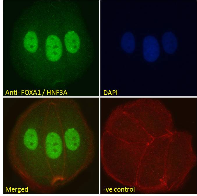
Immunocytochemistry - Anti-FOXA1 antibody (ab5089)
Immunofluorescent analysis of paraformaldehyde-fixed, 0.15% TritonX-100 permeabilized U-2 OS (Human bone osteosarcoma epithelial cell line) cells labeling FOXA1 with ab5089 at 10 ug/ml (1h). Image showing nuclear staining in U-2 OS cells. An Alexa Fluor 488® secondary antibody was used at 2 ug/ml. Actin filaments were stained with phalloidin (red) and the nuclear stain is DAPI (blue).
Secondary antibody only control: Unimmunized goat IgG (10 ug/ml) followed by an Alexa Fluor 488® secondary antibody at 2ug/ml.


