Anti-p38 (phospho T180 + Y182)抗体
参阅全部 p38 一抗
兔多克隆抗体to p38 (phospho T180 + Y182)
Rabbit
适用于: IHC-P, ICC/IF, WBmore details
与反应: Rat, Human
预测可用于: Mouse, Dog, Carp, Monkey![]()
Synthetic peptide corresponding to Human p38 (phospho T180 + Y182). p38 is dually phosphorylated and therefore fully activated by MEK3 and MEK6 on threonine 180 and tyrosine 182 within the activation loop.
Database link: Q16539
(Peptide available as ab5253)
WB: HeLa, A431, COLO 205, A549 and A549 cell lysate; HEK-293 (human epithelial cell line from embryonic kidney) cells. IHC-P: Human brain tissue, human heart tissue, rat heart tissue. ICC: SH-SY5Y (human neuroblastoma cell line from bone marrow) cells.
The Life Science industry has been in the grips of a reproducibility crisis for a number of years. Abcam is leading the way in addressing this with our range of recombinant monoclonal antibodies and knockout edited cell lines for gold-standard validation. Please check that this product meets your needs before purchasing.
If you have any questions, special requirements or concerns, please send us an inquiry and/or contact our Support team ahead of purchase. Recommended alternatives for this product can be found below, along with publications, customer reviews and Q&As
Liquid
Shipped at 4°C. Store at +4°C short term (1-2 weeks). Upon delivery aliquot. Store at -20°C. Avoid freeze / thaw cycle.
pH: 7.30
Preservative: 0.05% Sodium azide
Constituents: PBS, 50% Glycerol, 0.1% BSA
BSA is IgG and protease free. PBS without Mg2+ and Ca2+.
浓度
50 µl 浓度为 0.2 mg/ml
Protein A purified
Purified from rabbit serum by sequential epitope-specific chromatography. The antibody has been negatively preadsorbed using i) non-phosphopeptide corresponding to the site of phosphorylation to remove antibody that is reactive with non-phosphorylated p38, and ii) a JNK-derived peptide that is phosphorylated at threonine 183 and tyrosine 185. The final product is generated by affinity chromatography using a p38-derived peptide that is phosphorylated at threonine 180 and tyrosine 182.
多克隆
IgG
Abpromise™承诺保证使用ab4822于以下的经测试应用
“应用说明”部分 下显示的仅为推荐的起始稀释度;实际最佳的稀释度/浓度应由使用者检定。
| 应用 | Ab评论 | 说明 |
|---|---|---|
| IHC-P | 1/10 - 1/100. | |
| ICC/IF | 1/250. | |
| WB | (3) | 1/1000. Predicted molecular weight: 38 kDa. |
Entrez Gene: 1432 Human
Entrez Gene: 26416 Mouse
Omim: 600289 Human
SwissProt: Q16539 Human
SwissProt: P47811 Mouse
Unigene: 485233 Human
Unigene: 311337 Mouse
Unigene: 88085 Rat
CSAID Binding Protein 1 antibody
CSAID binding protein antibody
CSAID-binding protein antibody
Csaids binding protein antibody
CSBP 1 antibody
CSBP 2 antibody
CSBP antibody
CSBP1 antibody
CSBP2 antibody
CSPB1 antibody
Cytokine suppressive anti-inflammatory drug-binding protein antibody
EXIP antibody
MAP kinase 14 antibody
MAP kinase MXI2 antibody
MAP kinase p38 alpha antibody
MAPK 14 antibody
MAPK14 antibody
MAX interacting protein 2 antibody
MAX-interacting protein 2 antibody
Mitogen Activated Protein Kinase 14 antibody
Mitogen activated protein kinase p38 alpha antibody
Mitogen-activated protein kinase 14 antibody
Mitogen-activated protein kinase p38 alpha antibody
MK14_HUMAN antibody
Mxi 2 antibody
MXI2 antibody
p38 ALPHA antibody
p38 antibody
p38 MAP kinase antibody
p38 MAPK antibody
p38 mitogen activated protein kinase antibody
p38ALPHA antibody
p38alpha Exip antibody
PRKM14 antibody
PRKM15 antibody
RK antibody
SAPK2A antibody
Stress-activated protein kinase 2a antibody

Western blot - Anti-p38 (phospho T180 + Y182) antibody (ab4822)
All lanes : Anti-p38 (phospho T180 + Y182) antibody (ab4822) at 1/1000 dilution
Lane 1 : HeLa (human epithelial cell line from cervix adenocarcinoma) cell lysate
Lane 2 : HeLa (human epithelial cell line from cervix adenocarcinoma) exposed for 40 min with UV, cell lysate
Lane 3 : A431 (human epidermoid carcinoma cell line) cell lysate
Lane 4 : A431 (human epidermoid carcinoma cell line) exposed for 40 min with UV, cell lysate
Lane 5 : COLO 205 (human colon adenocarcinoma cell line) cell lysate
Lane 6 : COLO 205 (human colon adenocarcinoma cell line) exposed for 40 min with UV, cell lysate
Lane 7 : A549 (human lung carcinoma cell line) cell lysate
Lane 8 : A549 (human lung carcinoma cell line) exposed for 40 min with UV, cell lysate
Lysates/proteins at 20 µg per lane.
Secondary
All lanes : Goat anti-Rabbit IgG HRP at 1/5000 dilution
Developed using the ECL technique.
Predicted band size: 38 kDa

Immunohistochemistry (Formalin/PFA-fixed paraffin-embedded sections) - Anti-p38 (phospho T180 + Y182) antibody (ab4822)
Paraffin-embedded human brain tissue stained for p38 (phospho T180 + Y182) using ab4822 (right panel) at 1/100 dilution in immunohistochemical analysis followed by HRP-conjugated secondary antibody and DAB staining. Negative control (left panel) staining without primary antibody.

Immunohistochemistry (Formalin/PFA-fixed paraffin-embedded sections) - Anti-p38 (phospho T180 + Y182) antibody (ab4822)
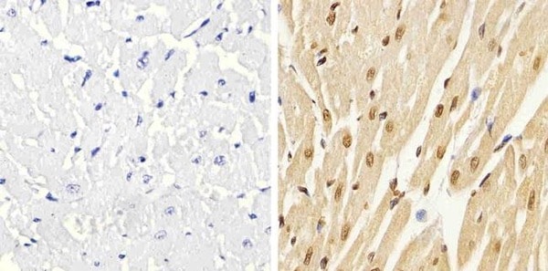
Paraffin-embedded human heart tissue stained for p38 (phospho T180 + Y182) using ab4822 (right panel) at 1/20 dilution in immunohistochemical analysis followed by HRP-conjugated secondary antibody and DAB staining. Negative control (left panel) staining without primary antibody.
Immunohistochemistry (Formalin/PFA-fixed paraffin-embedded sections) - Anti-p38 (phospho T180 + Y182) antibody (ab4822)
Paraffin-embedded rat heart tissue stained for p38 (phospho T180 + Y182) using ab4822 (right panel) at 1/20 dilution in immunohistochemical analysis followed by HRP-conjugated secondary antibody and DAB staining. Negative control (left panel) staining without primary antibody.

Immunocytochemistry/ Immunofluorescence - Anti-p38 (phospho T180 + Y182) antibody (ab4822)
4% PFA-fixed, Triton X-100 permeabilized SH-SY5Y (human neuroblastoma cell line from bone marrow) cells labeling p38 (phospho T180 + Y182) (Panel A: green) using ab4822 at 1 µg/mL in ICC/IF. Secondary antibody: Alexa Flour® 488 Goat Anti-Rabbit IgG at 1/400 dilution. Nuclei (Panel b: blue) were stained with SlowFade® Gold Antifade Mountant with DAPI. F-actin (Panel c: red) was stained with Alexa Fluor® 594 Phalloidin. Panel d is a merged image showing nuclear localization. Panel e is a no primary antibody control.

Western blot - Anti-p38 (phospho T180 + Y182) antibody (ab4822)
Peptide Competition: Extracts prepared from HEK-293 (human epithelial cell line from embryonic kidney) cells treated with UV irradiation were resolved on a 10% Tris-glycine gel and transferred to nitrocellulose. Membranes were blocked with a 5% BSA-TBST buffer overnight at 4oC, then were incubated with 0.50 µg/mL ab4822 for two hours at room temperature in a 3% BSA-TBST buffer, following its prior incubation with: the peptide immunogen (1), a generic phosphothreonine containing peptide (2), a generic phosphotyrosine-containing peptide (3), the non-phosphorylated peptide corresponding to the phosphopeptide (4), no peptide (5), the phosphorylated peptide derived from the corresponding region of JNK 1 & 2 (6), and, the phosphorylated peptide derived from the corresponding region of ERK 1 & 2 (7). After washing, membranes were incubated with goat F(ab')2 antirabbit IgG alkaline phosphatase and the signal was detected using the Tropix WesternStar method. The data show that only the phosphopeptide
抱歉,暂无浏览记录


