Anti-SORBS1抗体
参阅全部 SORBS1 一抗
山羊多克隆抗体to SORBS1
Goat
This antibody is expected to recognise both human isoforms.
适用于: WB, IHC-Pmore details
与反应: Human
预测可用于: Mouse, Rat![]()
Synthetic peptide corresponding to Human SORBS1 aa 800-900 (C terminal). (NP_056200.1; NP_001030126.1; NP_001030127.1; NP_001030128.1; NP_079267.1; NP_001030129.1)
Database link: NP_006425.2
The Life Science industry has been in the grips of a reproducibility crisis for a number of years. Abcam is leading the way in addressing this with our range of recombinant monoclonal antibodies and knockout edited cell lines for gold-standard validation. Please check that this product meets your needs before purchasing.
If you have any questions, special requirements or concerns, please send us an inquiry and/or contact our Support team ahead of purchase. Recommended alternatives for this product can be found below, along with publications, customer reviews and Q&As
Liquid
Shipped at 4°C. Upon delivery aliquot and store at -20°C or -80°C. Avoid repeated freeze / thaw cycles.
pH: 7.3
Preservative: 0.02% Sodium azide
Constituents: Tris buffered saline, 0.5% BSA
Immunogen affinity purified
Purified from goat serum by ammonium sulphate precipitation followed by antigen affinity chromatography using the immunizing peptide.
多克隆
IgG
Abpromise™承诺保证使用ab4551于以下的经测试应用
“应用说明”部分 下显示的仅为推荐的起始稀释度;实际最佳的稀释度/浓度应由使用者检定。
| 应用 | Ab评论 | 说明 |
|---|---|---|
| WB | (1) | Use at an assay dependent concentration. A 1 hour primary incubation is recommended for this product. No signal obtained yet but low background observed in human heart and 293 lysates at up to 2 µg/ml. |
| IHC-P | Use a concentration of 2.5 µg/ml. Perform heat mediated antigen retrieval before commencing with IHC staining protocol. |
Entrez Gene: 10580 Human
Entrez Gene: 20411 Mouse
Omim: 605264 Human
SwissProt: Q9BX66 Human
SwissProt: Q62417 Mouse
Unigene: 38621 Human
Unigene: 210815 Mouse
Unigene: 417719 Mouse
Unigene: 440351 Mouse
Unigene: 110441 Rat
c-Cbl-associated protein antibody
CAP antibody
CBL Associated Protein antibody
Ponsin antibody
SH3 domain protein 5 antibody
SH3D5 antibody
SH3P12 antibody
Sorbin and SH3 Domain Containing 1 antibody
Sorbin and SH3 domain-containing protein 1 antibody
Sorbs1 antibody
SRBS1_HUMAN antibody
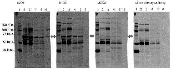
Western blot - Anti-SORBS1 antibody (ab4551)
All lanes : Anti-SORBS1 antibody (ab4551)
Lane 1 : Molecular weight marker (Bio-Rad Precision Plus Kaleidoscope standards)
Lane 2 : 10 µl Human plasma 1 (600-800 µg protein)
Lane 3 : 10 µl Human plasma 2 (600-800 µg protein)
Lane 4 : 10 µl Human White Blood cells 1 (30-50 µg protein)
Lane 5 : 10 µl Human White Blood cells 2 (30-50 µg protein)
Lane 6 : 10 µl HEK293 whole cell lysate (prepared by scrapping 10 cm confluent dish into 4ml of 2x sample buffer (Sigma)
Secondary
All lanes : Donkey anti-goat IgG (H+L) conjugated to Alexa-Fluor680 at 1/5000 dilution
ab4551 was tested in in human plasma, human white blood cells and HEK293 cells. Samples were run for 1h at 200 V using the Fisherbrand vertical complete gel unit, and transferred onto nitrocellulose 0.45um (1h, 100V, 4 C) using the Bio-rad Trans blot cell with plate electrodes. Membranes were blocked for 1 h at RT, then incubated overnight in primary antibody (1/200, 1/1000, 1/5000 or minus primary antibody) at 4 degrees C, with shaking. Following washing, membranes were incubated in secondary antibody (1/5000, Alexa-Fluor680 donkey anti-goat IgG, H+L) for 1 h at RT. Antibody binding was detected using LI-COR Bioscience's Odyssey Infrared Imaging system (intensity 5). The molecular weight of sorbs 1 was calculated using 'Compute pI/MW'from ExPASy tools based on the amino acid sequence according to NP_006425. The band indicated is approximately this predicted molecular weight, and is not present in the minus primary image, scanned at the same intensity.
Image courtesy of an anonymous abreview.

Immunohistochemistry (Formalin/PFA-fixed paraffin-embedded sections) - Anti-SORBS1 antibody (ab4551)
ab4551 (2.5 µg/ml) staining SORBS1 in Human Skeletal Muscle by IHC-P. Steamed antigen retrieval in citrate buffer pH6, AP-staining. This image shows spotty staining on muscle fibres in longitudinal section.
抱歉,暂无浏览记录


