Anti-NLRP3抗体
参阅全部 NLRP3 一抗
山羊多克隆抗体to NLRP3
Goat
适用于: Flow Cyt (Intra), WBmore details
不适用于: ICC/IF
与反应: Human
预测可用于: Rat, Cow![]()
Synthetic peptide corresponding to Human NLRP3 aa 1024-1036 (C terminal).
Sequence:
C-EKPELTVVFEPSW
Database link: Q96P20-1
WB: THP-1 cells Flow Cyt (intra): U937 cells.
No signal has been obtained in Western blot but low background has observed in Daudi, A431, Jurkat, U937 and MOLT-4 lysates at up to 1µg/ml. We would appreciate any feedback from people in the field.
The Life Science industry has been in the grips of a reproducibility crisis for a number of years. Abcam is leading the way in addressing this with our range of recombinant monoclonal antibodies and knockout edited cell lines for gold-standard validation. Please check that this product meets your needs before purchasing.
If you have any questions, special requirements or concerns, please send us an inquiry and/or contact our Support team ahead of purchase. Recommended alternatives for this product can be found below, along with publications, customer reviews and Q&As
Liquid
Shipped at 4°C. Upon delivery aliquot and store at -20°C or -80°C. Avoid repeated freeze / thaw cycles.
pH: 7.30
Preservative: 0.02% Sodium azide
Constituents: 0.5% BSA, Tris buffered saline
Immunogen affinity purified
Purified from goat serum by ammonium sulphate precipitation followed by antigen affinity chromatography using the immunizing peptide.
多克隆
IgG
Abpromise™承诺保证使用ab4207于以下的经测试应用
“应用说明”部分 下显示的仅为推荐的起始稀释度;实际最佳的稀释度/浓度应由使用者检定。
| 应用 | Ab评论 | 说明 |
|---|---|---|
| Flow Cyt (Intra) | Use a concentration of 10 µg/ml. ab37373 - Goat polyclonal IgG, is suitable for use as an isotype control with this antibody. | |
| WB | (2) | 1/1000. Predicted molecular weight: 118 kDa. |
应用说明
Is unsuitable for ICC/IF.
Entrez Gene: 114548 Human
Omim: 606416 Human
SwissProt: Q96P20 Human
Unigene: 159483 Human
Angiotensin/vasopressin receptor AII/AVP-like antibody
C1orf7 antibody
Caterpiller protein 1.1 antibody
CIAS 1 antibody
CIAS1 antibody
CLR1.1 antibody
Cold autoinflammatory syndrome 1 antibody
Cold autoinflammatory syndrome 1 protein antibody
Cryopyrin antibody
Familial cold autoinflammatory syndrome antibody
FCAS antibody
FCU antibody
LRR and PYD domains-containing protein 3 antibody
Muckle-Wells syndrome antibody
MWS antibody
NACHT antibody
NACHT LRR and PYD containing protein 3 antibody
NALP 3 antibody
NALP3 antibody
NALP3_HUMAN antibody
NLR family pyrin domain containing 3 antibody
NLRP3 antibody
PYPAF 1 antibody
PYPAF1 antibody
PYRIN containing APAF1 like protein 1 antibody
PYRIN-containing APAF1-like protein 1 antibody
AGTAVPRL antibody
AII/AVP antibody
Angiotensin/vasopressin receptor AII/AVP like antibody
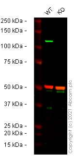
Western blot - Anti-NLRP3 antibody (ab4207)
All lanes : Anti-NLRP3 antibody (ab4207) at 1/1000 dilution
Lane 1 : Wild-type THP-1 cell lysate
Lane 2 : NLRP3 knockout THP-1 cell lysate
Lysates/proteins at 20 µg per lane.
Performed under reducing conditions.
Predicted band size: 118 kDa
Observed band size: 118 kDa
False colour image of Western blot: Anti-NLRP3 antibody staining at 1/1000 dilution, shown in green; Mouse anti-Alpha Tubulin [DM1A] (ab7291) loading control staining at 1/20000 dilution, shown in red. In Western blot, ab4207 was shown to bind specifically to NLRP3. A band was observed at 118 kDa in wild-type THP-1 cell lysates with no signal observed at this size in NLRP3 knockout cell line ab280063 (knockout cell lysate ab280122). To generate this image, wild-type and NLRP3 knockout THP-1 cell lysates were analysed. First, samples were run on an SDS-PAGE gel then transferred onto a nitrocellulose membrane. Membranes were blocked in fluorescent western blot (TBS-based) blocking solution before incubation with primary antibodies overnight at 4 °C. Blots were washed four times in TBS-T, incubated with secondary antibodies for 1 h at room temperature, washed again four times then imaged. Secondary antibodies used were Donkey anti-Goat IgG H&L (IRDye® 800CW) preabsorbed (ab216775) and Donkey anti-Mouse IgG H&L (IRDye® 680RD) preabsorbed (ab216778) at 1/20000 dilution.

Flow Cytometry (Intracellular) - Anti-NLRP3 antibody (ab4207)
Flow cytometric analysis of paraformaldehyde fixed U937 cells (blue line), permeabilized with 0.5% Triton. Primary incubation with ab4207 was 1hr (10ug/ml) followed by Alexa Fluor® 488 secondary antibody (1ug/ml). IgG control: Unimmunized goat IgG (black line) followed by Alexa Fluor® 488 secondary antibody.

Flow Cytometry (Intracellular) - Anti-NLRP3 antibody (ab4207)This image is courtesy of an Abreview submitted by Mahesh Shivananjappa.
ab4207 staining NLRP3 in the Human White Blood Cells (Mixed Population) by Flow Cytometry. WBC were isolated spinning Blood on Ficoll Gradient after removal of RBC's and permeabilized with 0.1% Triton-X100 in 2% BSA for 15 minutes. The sample was incubated with the primary antibody (1/100 in PBS + 2% BSA in PBS) for 16 hours at 4°C. An Alexa Flour® 488 Donkey Anti Goat IgG (H+L) (1/250) was used as the secondary antibody.
Gating Strategy: Monocytes


