Anti-PCSK2抗体
参阅全部 PCSK2 一抗
兔多克隆抗体to PCSK2
Rabbit
适用于: ICC/IF, WBmore details
与反应: Mouse, Human
预测可用于: Rat, Cow, Pig![]()
Synthetic peptide corresponding to Mouse PCSK2 aa 600-700.
The Life Science industry has been in the grips of a reproducibility crisis for a number of years. Abcam is leading the way in addressing this with our range of recombinant monoclonal antibodies and knockout edited cell lines for gold-standard validation. Please check that this product meets your needs before purchasing.
If you have any questions, special requirements or concerns, please send us an inquiry and/or contact our Support team ahead of purchase. Recommended alternatives for this product can be found below, along with publications, customer reviews and Q&As
Liquid
Shipped at 4°C. Store at +4°C short term (1-2 weeks). Upon delivery aliquot. Store at -20°C or -80°C. Avoid freeze / thaw cycle.
Preservative: 0.05% Sodium azide
Constituents: 0.1% BSA, 99% PBS
浓度
100 µg 浓度为 1 mg/ml
Immunogen affinity purified
The subtilisin-like Prohormone Convertase (PC) family is a group of cellular enzymes that cleave most prohormones and neuropeptide precursors. Numerous other cellular proteins, some viral proteins, and bacterial toxins that are transported by the constitutive secretory pathway are also targeted for maturation by PCs. PC family members share structural similarities, which include a heterogeneous ~10 kDa amino-terminal proregion, a highly conserved ~55 kDa subtilisin-like catalytic domain, and carboxyl-terminal domain that is heterogeneous in length and sequence. These enzymes become catalytically active following proregion cleavage within the appropriate cellular compartment. The subcellular localization of PC family members varies. Immunolocalization studies show that PC1 is found in the perinuclear region as well as the trans-Golgi network, whereas PC2 can be found in the trans-Golgi network as well as diffusely distributed in the peripheral cytoplasm.
多克隆
IgG
Abpromise™承诺保证使用ab3533于以下的经测试应用
“应用说明”部分 下显示的仅为推荐的起始稀释度;实际最佳的稀释度/浓度应由使用者检定。
| 应用 | Ab评论 | 说明 |
|---|---|---|
| ICC/IF | 1/20 - 1/200. | |
| WB | Use a concentration of 2 µg/ml. This antibody detects an ~65 kDa protein representing Proprotein 2 from alpha TC 6 cell extract. |
Entrez Gene: 5126 Human
Entrez Gene: 18549 Mouse
Omim: 162151 Human
SwissProt: P16519 Human
SwissProt: P21661 Mouse
Unigene: 315186 Human
Unigene: 294493 Mouse
Unigene: 447466 Mouse
Unigene: 89052 Rat
KEX2 like endoprotease 2 antibody
KEX2-like endoprotease 2 antibody
NEC 2 antibody
NEC2 antibody
NEC2_HUMAN antibody
Neuroendocrine convertase 2 antibody
PC2 antibody
PCSK2 antibody
Prohormone convertase 2 antibody
Proprotein convertase 2 antibody
Proprotein convertase subtilisin/kexin type 2 antibody
SPC2 antibody

Western blot - Anti-PCSK2 antibody (ab3533)
Anti-PCSK2 antibody (ab3533) at 1/500 dilution + Mouse cerebellum cell lysate at 25 µg
Observed band size: 65 kDawhy is the actual band size different from the predicted?
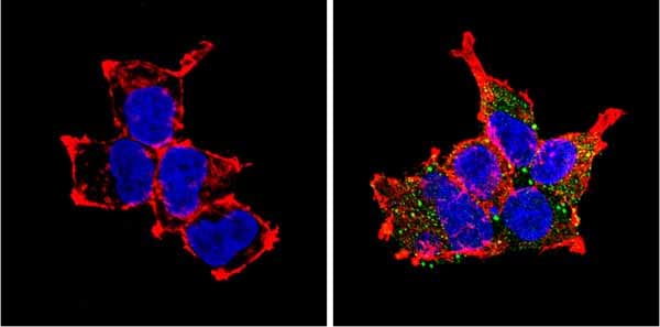
Immunocytochemistry/ Immunofluorescence - Anti-PCSK2 antibody (ab3533)
Immunocytochemistry/Immunofluorescent analysis of PCSK2 (green) showing staining in the cytoplasm of HEK293 cells. Formalin-fixed cells were permeabilized with 0.1% Triton X-100 in TBS for 5-10 minutes and blocked with 3% BSA-PBS for 30 minutes at room temperature. Cells were probed with ab3533 in 3% BSA-PBS at a dilution of 1:100 and incubated overnight at 4°C in a humidified chamber. Cells were washed with PBST and incubated with a DyLight-conjugated secondary antibody in PBS at room temperature in the dark. F-actin (red) was stained with a fluorescent red phalloidin and nuclei (blue) were stained with Hoechst or DAPI. Images were taken at a magnification of 60x.

Immunocytochemistry/ Immunofluorescence - Anti-PCSK2 antibody (ab3533)
Immunocytochemistry/Immunofluorescent analysis of PCSK2 (green) showing staining in the cytoplasm of C2C12 cells. Formalin-fixed cells were permeabilized with 0.1% Triton X-100 in TBS for 5-10 minutes and blocked with 3% BSA-PBS for 30 minutes at room temperature. Cells were probed with ab3533 in 3% BSA-PBS at a dilution of 1:100 and incubated overnight at 4°C in a humidified chamber. Cells were washed with PBST and incubated with a DyLight-conjugated secondary antibody in PBS at room temperature in the dark. F-actin (red) was stained with a fluorescent red phalloidin and nuclei (blue) were stained with Hoechst or DAPI. Images were taken at a magnification of 60x.
抱歉,暂无浏览记录


