Anti-Adiponectin抗体
参阅全部 Adiponectin 一抗
兔多克隆抗体to Adiponectin
Rabbit
Detects Acrp30 from mouse serum. We have data to indicate that this antibody may not cross react with Human. However, this has not been conclusively tested and expression levels may vary in certain cell lines/tissues.
适用于: ICC, WBmore details
与反应: Mouse
This product was produced with the following immunogens:
Synthetic peptide corresponding to Mouse Adiponectin aa 1-100.
Database link: Q60994
Synthetic peptide corresponding to Mouse Adiponectin aa 150-250.
Database link: Q60994
The Life Science industry has been in the grips of a reproducibility crisis for a number of years. Abcam is leading the way in addressing this with our range of recombinant monoclonal antibodies and knockout edited cell lines for gold-standard validation. Please check that this product meets your needs before purchasing.
If you have any questions, special requirements or concerns, please send us an inquiry and/or contact our Support team ahead of purchase. Recommended alternatives for this product can be found below, along with publications, customer reviews and Q&As
Liquid
Shipped at 4°C. Store at +4°C short term (1-2 weeks). Upon delivery aliquot. Store at -20°C or -80°C. Avoid freeze / thaw cycle.
Preservative: 0.05% Sodium azide
Constituents: 0.1% BSA, 99% PBS
Protein A purified
多克隆
IgG
Abpromise™承诺保证使用ab3455于以下的经测试应用
“应用说明”部分 下显示的仅为推荐的起始稀释度;实际最佳的稀释度/浓度应由使用者检定。
| 应用 | Ab评论 | 说明 |
|---|---|---|
| ICC | Use a concentration of 2 µg/ml. | |
| WB | Use a concentration of 4 - 8 µg/ml. Detects a band of approximately 30 kDa. |
Entrez Gene: 11450 Mouse
SwissProt: Q60994 Mouse
Unigene: 3969 Mouse
Acrp 30 antibody
ACRP30 antibody
ADIPO_HUMAN antibody
Adipocyte antibody
Adipocyte C1q and collagen domain containing protein antibody
Adipocyte complement related 30 kDa protein antibody
Adipocyte complement related protein of 30 kDa antibody
Adipocyte complement-related 30 kDa protein antibody
adipocyte-specific secretory protein antibody
Adiponectin antibody
Adiponectin precursor antibody
adiponectin, C1Q and collagen domain containing antibody
Adipoq antibody
Adipose most abundant gene transcript 1 antibody
Adipose most abundant gene transcript 1 protein antibody
Adipose specific collagen like factor antibody
ADIPQTL1 antibody
ADPN antibody
APM 1 antibody
apM-1 antibody
APM1 antibody
C1q and collagen domain-containing protein antibody
GBP 28 antibody
GBP28 antibody
Gelatin binding protein antibody
Gelatin binding protein 28 antibody
gelatin-binding protein 28 antibody
Gelatin-binding protein antibody
OTTHUMP00000210047 antibody
30 kDa adipocyte complement related protein antibody
30 kDa adipocyte complement-related protein antibody
ACDC antibody

Western blot - Anti-Adiponectin antibody (ab3455)
All lanes : Anti-Adiponectin antibody (ab3455) at 2 µg/ml
Lane 1 : Mouse adipose tissue extract in 3T3-L1 conditioned media
Lane 2 : Mouse adipose tissue extract in NIH/3T3 conditioned media
Lane 3 : Mouse adipose tissue extract in BM-MSC differentiated conditioned media
Lysates/proteins at 30 µg per lane.
Secondary
All lanes : Goat anti-Rabbit IgG (H+L) Superclonal™ Secondary Antibody, HRP conjugate at 1/4000 dilution
Developed using the ECL technique.
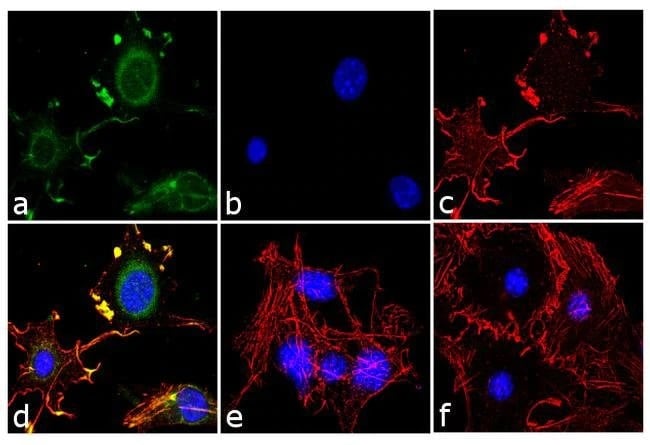
Immunocytochemistry - Anti-Adiponectin antibody (ab3455)
Immunofluorescence analysis of Adiponectin was performed using 70% confluent 3T3-L1 cells differentiated with Adipogenesis Assay Kit (Cell-Based)(ab133102) for 5 days. The cells were fixed with 4% paraformaldehyde for 10 minutes, permeabilized with 0.1% Triton TM X-100 for 10 min, blocked with 1% BSA for 1 hour at room temperature.
The cells were labeled with Adiponectin Polyclonal Antibody (ab3455) at 2 μg/ml in 0.1% BSA and incubated for 3 hours at room temperature and then lebeled with Goat Anti-Rabbit IgG H&L (Alexa Fluor® 488) preadsorbed (ab150081) at a dilution of 1/2000 for 45 minutes at room temperature, panel A (green). Panel B (blue): nuclei were stained with DAPI. Panel C (red), F-actin was stained with Alexa Fluor®555 rhodamine phalloidin (ab235138) at 1/3000 dilution. Panel D represents the merged image showing cytoplasmic localization. Panel f represents control cells with no primary antibody to assess background. The images were captured at 60X magnification.

Western blot - Anti-Adiponectin antibody (ab3455)
Western blot detection of Adiponectin on mouse serum using ab3455.
抱歉,暂无浏览记录


