Anti-Munc18-1抗体
参阅全部 Munc18-1 一抗
兔多克隆抗体to Munc18-1
Rabbit
适用于: WB, ICCmore details
与反应: Mouse, Rat, Human
预测可用于: Chicken, Cow, Pig![]()
Synthetic peptide corresponding to Human Munc18-1 aa 58-70.
Sequence:
VEDINKRREPIPS
(Peptide available as ab4982)
WB: Mouse and rat brain tissue lysates ICC: U-87 MG cells
The Life Science industry has been in the grips of a reproducibility crisis for a number of years. Abcam is leading the way in addressing this with our range of recombinant monoclonal antibodies and knockout edited cell lines for gold-standard validation. Please check that this product meets your needs before purchasing.
If you have any questions, special requirements or concerns, please send us an inquiry and/or contact our Support team ahead of purchase. Recommended alternatives for this product can be found below, along with publications, customer reviews and Q&As
Liquid
Shipped at 4°C. Store at +4°C short term (1-2 weeks). Upon delivery aliquot. Store at -20°C or -80°C. Avoid freeze / thaw cycle.
Preservative: 0.05% Sodium azide
Constituents: 0.1% BSA, 99% PBS
浓度
100 µg 浓度为 1 mg/ml
Immunogen affinity purified
多克隆
IgG
Abpromise™承诺保证使用ab3451于以下的经测试应用
“应用说明”部分 下显示的仅为推荐的起始稀释度;实际最佳的稀释度/浓度应由使用者检定。
| 应用 | Ab评论 | 说明 |
|---|---|---|
| WB | (2) | Use a concentration of 1 µg/ml. Detects a band of approximately 67 kDa. By Western blot, this antibody detects an ~67 kDa protein representing Munc-18 from rat whole brain extract. |
| ICC | Use a concentration of 2 µg/ml. |
Entrez Gene: 404293 Chicken
Entrez Gene: 6812 Human
Entrez Gene: 20910 Mouse
Omim: 602926 Human
SwissProt: Q6R748 Chicken
SwissProt: P61764 Human
SwissProt: O08599 Mouse
Unigene: 288229 Human
Unigene: 278865 Mouse
Unigene: 80843 Rat
FLJ37475 antibody
Munc 18 1 antibody
Munc 18a antibody
MUNC18 1 antibody
N-Sec1 antibody
Neuronal SEC1 antibody
NSec1 antibody
p67 antibody
Protein unc-18 homolog 1 antibody
Protein unc-18 homolog A antibody
Rb sec1 antibody
RBSEC1 antibody
STXB1_HUMAN antibody
STXBP1 antibody
Syntaxin binding protein 1 antibody
Syntaxin-binding protein 1 antibody
Unc 18 homolog antibody
Unc 18A antibody
Unc-18A antibody
Unc18 1 antibody
UNC18 antibody
Unc18-1 antibody

Western blot - Anti-Munc18-1 antibody (ab3451)
Anti-Munc18-1 antibody (ab3451) at 2 µg/ml + Mouse brain tissue lysate at 30 µg
Secondary
Goat anti-Rabbit IgG (H+L) Superclonal™ at 1/4000 dilution
Observed band size: 67 kDawhy is the actual band size different from the predicted?
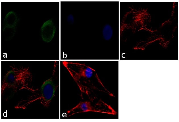
Immunocytochemistry - Anti-Munc18-1 antibody (ab3451)
Immunocytochemistry analysis of 70% confluent log phase U-87 MG cells labeling Munc18-1 with ab3451 at 2µg/mL in 0.1% BSA and incubated for 3 hours at room temperature followed by Goat anti-Rabbit IgG (H+L) Superclonal™ Secondary Antibody, Alexa Fluor® 488 conjugate at a dilution of 1/2000 for 45 minutes at room temperature (Panel a: green). The cells were fixed with 4% paraformaldehyde for 10 minutes, permeabilized with 0.1% Triton™ X-100 for 10 minutes, and blocked with 1% BSA for 1 hour at room temperature. Nuclei (Panel b: blue) were stained with SlowFade® Gold Antifade Mountant with DAPI. F-actin (Panel c: red) was stained with Alexa Fluor® 555 Rhodamine Phalloidin. Panel d represents the merged image showing cytoplasmic localization. Panel e shows the no primary antibody control. The images were captured at 60X magnification.

Western blot - Anti-Munc18-1 antibody (ab3451)
All lanes : Anti-Munc18-1 antibody (ab3451) at 2 µg/ml
Lane 1 : Mouse brain tissue lysate
Lane 2 : Rat brain tissue lysate
Lane 3 : Rat liver tissue lysate
Lane 4 : Rat kidney tissue lysate
Lysates/proteins at 30 µg per lane.
Secondary
All lanes : Goat anti-Rabbit IgG (H+L) Superclonal™ at 1/4000 dilution
Observed band size: 67 kDawhy is the actual band size different from the predicted?
Detected by chemiluminescence

Western blot - Anti-Munc18-1 antibody (ab3451)This image is courtesy of an anonymous Abreview
Anti-Munc18-1 antibody (ab3451) at 1/5000 dilution + Mouse hippocampal homogenate at 5 µg
Secondary
Goat Anti-Rabbit IgG H&L (HRP) preadsorbed (ab97069) at 1/10000 dilution
Developed using the ECL technique.
Performed under reducing conditions.
Observed band size: 67 kDawhy is the actual band size different from the predicted?
Exposure time: 1 minute

Western blot - Anti-Munc18-1 antibody (ab3451)
Western blot of Munc-18 in rat brain homogenate immunoprecipitated with ab3451.
1. Extract only.
2-4. 750 ug of extract immunoprecipitated with 1 ug, 5 ug, and 10 ug of ab3451, respectively.
抱歉,暂无浏览记录


