Anti-NFAT5抗体
参阅全部 NFAT5 一抗
兔多克隆抗体to NFAT5
Rabbit
Detects Nuclear Factor of Activated T-cells 5 (NFAT 5).
适用于: IHC-P, ICC/IF, WB, IPmore details
与反应: Mouse, Human, African green monkey
Synthetic peptide corresponding to Human NFAT5 aa 1439-1455 (C terminal).
Sequence:
DLLVSLQNQGNNLTGSF
(Peptide available as ab4978)
WB: MCF7, Jurkat, Raji, Ramos, HepG2, U2OS, HeLa, COS7, EL4, C2C12 and NRK cell lysates. IHC-P: Human brain and skeletal muscle tissues. ICC/IF: NIH/3T3, MCF7 and HeLa cells. IP: U2OS cells.
The Life Science industry has been in the grips of a reproducibility crisis for a number of years. Abcam is leading the way in addressing this with our range of recombinant monoclonal antibodies and knockout edited cell lines for gold-standard validation. Please check that this product meets your needs before purchasing.
If you have any questions, special requirements or concerns, please send us an inquiry and/or contact our Support team ahead of purchase. Recommended alternatives for this product can be found below, along with publications, customer reviews and Q&As
Liquid
Shipped at 4°C. Store at +4°C short term (1-2 weeks). Upon delivery aliquot. Store at -20°C or -80°C. Avoid freeze / thaw cycle.
Preservative: 0.05% Sodium azide
Constituents: 0.1% BSA, 99% PBS
浓度
50 µg 浓度为 1 mg/ml
Immunogen affinity purified
多克隆
IgG
Abpromise™承诺保证使用ab3446于以下的经测试应用
“应用说明”部分 下显示的仅为推荐的起始稀释度;实际最佳的稀释度/浓度应由使用者检定。
| 应用 | Ab评论 | 说明 |
|---|---|---|
| IHC-P | 1/20. Perform heat mediated antigen retrieval with citrate buffer pH 6 before commencing with IHC staining protocol. | |
| ICC/IF | 1/100. | |
| WB | (1) | 1/1000. Detects a band of approximately 170 kDa (predicted molecular weight: 160 kDa).Can be blocked with NFAT5 peptide (ab4978). |
| IP | Use at an assay dependent concentration. 3 μg |
Entrez Gene: 10725 Human
Entrez Gene: 54446 Mouse
Omim: 604708 Human
SwissProt: O94916 Human
SwissProt: Q9WV30 Mouse
Unigene: 371987 Human
Unigene: 390057 Mouse
NF-AT5 antibody
NFAT 5 antibody
NFAT L1 antibody
NFAT like protein 1 antibody
NFAT5 antibody
NFAT5_HUMAN antibody
NFATL 1 antibody
NFATL1 antibody
NFATZ antibody
Nuclear factor of activated T cells 5 antibody
Nuclear factor of activated T cells 5 tonicity responsive antibody
Nuclear factor of activated T cells antibody
Nuclear factor of activated T-cells 5 antibody
OREBP antibody
Osmotic response element binding protein antibody
T cell transcription factor NFAT 5 antibody
T cell transcription factor NFAT5 antibody
T-cell transcription factor NFAT5 antibody
TonE binding protein antibody
TonE-binding protein antibody
TonEBP antibody
Tonicity responsive enhancer binding protein antibody
Tonicity-responsive enhancer-binding protein antibody
Glutamine rich protein H65 antibody
KIAA0827 antibody
NF AT5 antibody

Western blot - Anti-NFAT5 antibody (ab3446)
All lanes : Anti-NFAT5 antibody (ab3446) at 1/1000 dilution
Lane 1 : MCF7 (Human breast adenocarcinoma cell line) whole cell lysate
Lane 2 : Jurkat (Human T cell leukemia cell line from peripheral blood) whole cell lysate
Lane 3 : Raji (Human Burkitt's lymphoma cell line) whole cell lysate
Lane 4 : Ramos (Human Burkitt's lymphoma cell line) whole cell lysate
Lane 5 : HepG2 (Human liver hepatocellular carcinoma cell line) whole cell lysate
Lane 6 : U-2 OS (Human bone osteosarcoma epithelial cell line) whole cell lysate
Lane 7 : HeLa (Human epithelial adenocarcinoma cell line) whole cell lysate
Lane 8 : COS-7 (African green monkey kidney fibroblast-like cell line) whole cell lysate
Lane 9 : EL4 (Mouse thymic lymphoma cell line) whole cell lysate
Lane 10 : C2C12 (Mouse myoblast cell line) whole cell lysate
Lane 11 : NRK (Rat kidney normal tissue) whole cell lysate
Lysates/proteins at 25 µg per lane.
Secondary
All lanes : Goat anti-rabbit-HRP secondary antibody at 1/20000 dilution
Predicted band size: 160 kDa
Western blot analysis of NFAT5 was performed by loading samples onto a 4-20% Tris-HCl polyacrylamide gel. Proteins were transferred to a PVDF membrane and blocked with 5% Milk/TBST for at least 1 hour. Membranes were incubated with ab3446 overnight at 4°C on a rocking platform. Membranes were washed in TBS-0.1%Tween 20 and probed with a secondary antibody for at least one hour. Membranes were washed and chemiluminescent detection performed.
Immunocytochemistry/ Immunofluorescence - Anti-NFAT5 antibody (ab3446)
ICC analysis of NFAT5 in HeLa (Human epithelial adenocarcinoma cell line) cells. Cells were grown on chamber slides and fixed with formaldehyde prior to staining. Cells were probed without (control - right) or with ab3446 at a dilution of 1:20 overnight at 4 C, washed with PBS and incubated with a DyLight-488 conjugated secondary antibody. NFAT5 staining (green), F-Actin staining with Phalloidin (red) and nuclei with DAPI (blue) is shown. Images were taken at 60X magnification.

Immunohistochemistry (Formalin/PFA-fixed paraffin-embedded sections) - Anti-NFAT5 antibody (ab3446)
Immunohistochemistry was performed on normal biopsies of deparaffinized human skeletal muscle tissue. To expose target proteins, heat induced antigen retrieval was performed using 10mM sodium citrate (pH6.0) buffer, microwaved for 8-15 minutes. Following antigen retrieval tissues were blocked in 3% BSA-PBS for 30 minutes at room temperature. Tissues were then probed at a dilution of 1/20 duution with ab3446 or without primary antibody (negative control) overnight at 4°C in a humidified chamber. Tissues were washed extensively with PBST and endogenous peroxidase activity was quenched with a peroxidase suppressor. Detection was performed using a biotin-conjugated secondary antibody and SA-HRP, followed by colorimetric detection using DAB. Tissues were counterstained with hematoxylin and prepped for mounting.

Immunoprecipitation - Anti-NFAT5 antibody (ab3446)
Immunoprecipitation of NFAT5 was performed on U-2 OS (Human bone osteosarcoma epithelial cell line) whole cell lysate (lane 2). The antigen:antibody complex was formed by incubating 500 µg whole cell lysate with 3 µg of ab3446 overnight on a rocking platform at 4°C. The immune-complex was captured on 50 µL Protein A/G Plus Agarose. Captured immune-complexes were washed and proteins eluted with 5X Reducing Sample Loading Dye. Samples were resolved on a 4-20% Tris-HCl polyacrylamide gel. Proteins were transferred to PVDF membrane and blocked with 5% Milk/TBS-0.1%Tween for at least 1 hour. Membranes were washed in TBS-0.1%Tween 20 and probed with a goat anti-rabbit-HRP secondary antibody at a dilution of 1/20,000 for at least one hour. Membranes were washed and chemiluminescent detection performed.
Lane 1: Only cell lysate.

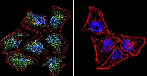
Immunocytochemistry/ Immunofluorescence - Anti-NFAT5 antibody (ab3446)
ICC analysis of NFAT5 in NIH/3T3 (Mouse embryo fibroblast cell line) cells. Cells were grown on chamber slides and fixed with formaldehyde prior to staining. Cells were probed without (control - right) or with ab3446 at a dilution of 1:20 overnight at 4 C, washed with PBS and incubated with a DyLight-488 conjugated secondary antibody. NFAT5 staining (green), F-Actin staining with Phalloidin (red) and nuclei with DAPI (blue) is shown. Images were taken at 60X magnification.

Immunohistochemistry (Formalin/PFA-fixed paraffin-embedded sections) - Anti-NFAT5 antibody (ab3446)
Immunohistochemistry was performed on normal biopsies of deparaffinized human brain tissue. To expose target proteins, heat induced antigen retrieval was performed using 10mM sodium citrate (pH6.0) buffer, microwaved for 8-15 minutes. Following antigen retrieval tissues were blocked in 3% BSA-PBS for 30 minutes at room temperature. Tissues were then probed at a dilution of 1:20 with ab3446 or without primary antibody (negative control) overnight at 4°C in a humidified chamber. Tissues were washed extensively with PBST and endogenous peroxidase activity was quenched with a peroxidase suppressor. Detection was performed using a biotin-conjugated secondary antibody and SA-HRP, followed by colorimetric detection using DAB. Tissues were counterstained with hematoxylin and prepped for mounting.
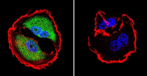
Immunocytochemistry/ Immunofluorescence - Anti-NFAT5 antibody (ab3446)
ICC analysis of NFAT5 in MCF7 (Human breast adenocarcinoma cell line) cells. Cells were grown on chamber slides and fixed with formaldehyde prior to staining. Cells were probed without (control - right) or with ab3446 at a dilution of 1:200 overnight at 4 C, washed with PBS and incubated with a DyLight-488 conjugated secondary antibody. NFAT5 staining (green), F-Actin staining with Phalloidin (red) and nuclei with DAPI (blue) is shown. Images were taken at 60X magnification.

Immunocytochemistry/ Immunofluorescence - Anti-NFAT5 antibody (ab3446)
ICC analysis of NFAT5 usingab3446 (shown in green) in HeLa (Human epithelial adenocarcinoma cell line) whole cells. Formalin fixed cells were permeabilized with 0.1% Triton X-100 in TBS for 10 minutes at room temperature. Cells were then blocked with 1% Blocker BSA for 15 minutes at room temperature. Cells were probed with a rabbit polyclonal antibody recognizing NFAT5, at a dilution of 1/100 for at least 1 hour at room temperature. Cells were washed with PBS and incubated with DyLight 488 goat-anti-rabbit secondary antibody at a dilution of 1/400 for 30 minutes at room temperature. Nuclei (blue) were stained with Hoechst 33342 dye. Images were taken on a Thermo Scientific ArrayScan at 20X magnification.


