Anti-Ubiquilin/UBQLN1抗体
兔多克隆抗体to Ubiquilin/UBQLN1
Rabbit
适用于: IHC-P, WBmore details
与反应: Mouse, Human, Chinese hamster
Synthetic peptide corresponding to Mouse Ubiquilin/UBQLN1 aa 2-18.
Sequence:
AESAESGGPPGAQDSAA
The Life Science industry has been in the grips of a reproducibility crisis for a number of years. Abcam is leading the way in addressing this with our range of recombinant monoclonal antibodies and knockout edited cell lines for gold-standard validation. Please check that this product meets your needs before purchasing.
If you have any questions, special requirements or concerns, please send us an inquiry and/or contact our Support team ahead of purchase. Recommended alternatives for this product can be found below, along with publications, customer reviews and Q&As
Liquid
Shipped at 4°C. Store at +4°C short term (1-2 weeks). Upon delivery aliquot. Store at -20°C or -80°C. Avoid freeze / thaw cycle.
Constituents: 0.1% BSA, 99% PBS
浓度
100 µg 浓度为 1 mg/ml
Immunogen affinity purified
多克隆
IgG
Abpromise™承诺保证使用ab3341于以下的经测试应用
“应用说明”部分 下显示的仅为推荐的起始稀释度;实际最佳的稀释度/浓度应由使用者检定。
| 应用 | Ab评论 | 说明 |
|---|---|---|
| IHC-P | 1/20 - 1/100. Perform heat mediated antigen retrieval with citrate buffer pH 6 before commencing with IHC staining protocol. | |
| WB | (1) | Use a concentration of 1 µg/ml. Predicted molecular weight: 63 kDa. By Western blot, this antibody detects an ~63 kDa protein representing Ubiquilin from CHO cell extract. |
Entrez Gene: 29979 Human
Entrez Gene: 56085 Mouse
Omim: 605046 Human
SwissProt: Q9UMX0 Human
SwissProt: Q8R317 Mouse
Unigene: 9589 Human
Unigene: 182053 Mouse
hPLIC-1 antibody
hPLIC1 antibody
PLIC-1 antibody
PLIC1 antibody
Protein linking IAP with cytoskeleton 1 antibody
Ubiquilin-1 antibody
Ubiquilin1 antibody
UBQL1_HUMAN antibody
UBQLN1 antibody
UBQN antibody
XDRP1 antibody
DA41 antibody
DSK2 antibody
FLJ90054 antibody

Immunohistochemistry (Formalin/PFA-fixed paraffin-embedded sections) - Anti-Ubiquilin/UBQLN1 antibody (ab3341)
Immunohistochemistry analysis of Ubiquilin showing staining in the nucleus and weak staining in the cytoplasm of paraffin-embedded human brain tissue (right) compared to a negative control without primary antibody (left). To expose target proteins, antigen retrieval was performed using 10mM sodium citrate (pH 6.0), microwaved for 8-15 min. Following antigen retrieval, tissues were blocked in 3% H2O2-methanol for 15 min at room temperature, washed with ddH2O and PBS, and then probed with ab3341 diluted in 3% BSA-PBS at a dilution of 1:100 for 1 hour at 37ºC in a humidified chamber. Tissues were washed extensively in PBST and detection was performed using an HRP-conjugated secondary antibody followed by colorimetric detection using a DAB kit. Tissues were counterstained with hematoxylin and dehydrated with ethanol and xylene to prep for mounting.

Immunohistochemistry (Formalin/PFA-fixed paraffin-embedded sections) - Anti-Ubiquilin/UBQLN1 antibody (ab3341)
Immunohistochemistry analysis of Ubiquilin showing staining in the cytoplasm and nucleus of paraffin-embedded human kidney tissue (right) compared to a negative control without primary antibody (left). To expose target proteins, antigen retrieval was performed using 10mM sodium citrate (pH 6.0), microwaved for 8-15 min. Following antigen retrieval, tissues were blocked in 3% H2O2-methanol for 15 min at room temperature, washed with ddH2O and PBS, and then probed with ab3341 diluted in 3% BSA-PBS at a dilution of 1:100 for 1 hour at 37ºC in a humidified chamber. Tissues were washed extensively in PBST and detection was performed using an HRP-conjugated secondary antibody followed by colorimetric detection using a DAB kit. Tissues were counterstained with hematoxylin and dehydrated with ethanol and xylene to prep for mounting.
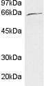
Western blot - Anti-Ubiquilin/UBQLN1 antibody (ab3341)
Anti-Ubiquilin/UBQLN1 antibody (ab3341) + CHO (Chinese hamster ovary cell line) whole cell lysate
Predicted band size: 63 kDa

Immunohistochemistry (Formalin/PFA-fixed paraffin-embedded sections) - Anti-Ubiquilin/UBQLN1 antibody (ab3341)
Immunohistochemistry analysis of Ubiquilin showing staining in the cytoplasm and nucleus of paraffin-embedded mouse kidney tissue (right) compared to a negative control without primary antibody (left). To expose target proteins, antigen retrieval was performed using 10mM sodium citrate (pH 6.0), microwaved for 8-15 min. Following antigen retrieval, tissues were blocked in 3% H2O2-methanol for 15 min at room temperature, washed with ddH2O and PBS, and then probed with ab3341 diluted in 3% BSA-PBS at a dilution of 1:20 for 1 hour at 37ºC in a humidified chamber. Tissues were washed extensively in PBST and detection was performed using an HRP-conjugated secondary antibody followed by colorimetric detection using a DAB kit. Tissues were counterstained with hematoxylin and dehydrated with ethanol and xylene to prep for mounting.


