Anti-PSMB5/MB1抗体
参阅全部 PSMB5/MB1 一抗
兔多克隆抗体to PSMB5/MB1
Rabbit
Detects proteasome PSMB5/MB1.
适用于: WB, ICCmore details
与反应: Mouse, Cow, Human
Synthetic peptide corresponding to Mouse PSMB5/MB1 aa 200 to the C-terminus.
The Life Science industry has been in the grips of a reproducibility crisis for a number of years. Abcam is leading the way in addressing this with our range of recombinant monoclonal antibodies and knockout edited cell lines for gold-standard validation. Please check that this product meets your needs before purchasing.
If you have any questions, special requirements or concerns, please send us an inquiry and/or contact our Support team ahead of purchase. Recommended alternatives for this product can be found below, along with publications, customer reviews and Q&As
Liquid
Shipped at 4°C. Store at +4°C short term (1-2 weeks). Upon delivery aliquot. Store at -20°C or -80°C. Avoid freeze / thaw cycle.
Constituents: 0.1% BSA, 99% PBS
浓度
100 µg 浓度为 1 mg/ml
Immunogen affinity purified
多克隆
IgG
Abpromise™承诺保证使用ab3330于以下的经测试应用
“应用说明”部分 下显示的仅为推荐的起始稀释度;实际最佳的稀释度/浓度应由使用者检定。
| 应用 | Ab评论 | 说明 |
|---|---|---|
| WB | (4) | 1/500 - 1/5000. Detects a band of approximately 22 kDa. |
| ICC | 1/10 - 1/200. |
Entrez Gene: 5693 Human
Entrez Gene: 19173 Mouse
Omim: 600306 Human
SwissProt: P28074 Human
SwissProt: O55234 Mouse
Unigene: 422990 Human
Unigene: 8911 Mouse
DKFZp459C139 antibody
EC 3.4.25.1 antibody
LMPX antibody
Macropain epsilon chain antibody
MB1 antibody
MGC104214 antibody
MGC118075 antibody
MGC134464 antibody
Multicatalytic endopeptidase complex epsilon chain antibody
Proteasome (prosome, macropain) subunit, beta type, 5 antibody
Proteasome beta 5 subunit antibody
Proteasome catalytic subunit 3 antibody
Proteasome chain 6 antibody
Proteasome epsilon chain antibody
Proteasome subunit beta type-5 antibody
Proteasome subunit MB1 antibody
Proteasome subunit X antibody
Proteasome subunit, beta type, 5 antibody
Proteasome subunit, beta-5 antibody
PSB5_HUMAN antibody
PSMB5 antibody
PSX large multifunctional protease X antibody
X antibody

Western blot - Anti-PSMB5/MB1 antibody (ab3330)
All lanes : Anti-PSMB5/MB1 antibody (ab3330) at 1/1000 dilution
Lane 1 : NCI-H1299 (Human lung carcinoma cell line) whole cell lysate
Lane 2 : HeLa (Human epithelial adenocarcinoma cell line) whole cell lysate
Lane 3 : Mouse liver cell lysate
Lysates/proteins at 25 µg per lane.
Observed band size: 23 kDawhy is the actual band size different from the predicted?
Immunocytochemistry - Anti-PSMB5/MB1 antibody (ab3330)
Immunocytochemistry/Immunofluorescence analysis of PSMB5/MB1 (green) showing staining in the cytoplasm and nucleus of A431 (Human epidermoid carcinoma cell line) cells (right) compared to a negative control without primary antibody (left). Formalin-fixed cells were permeabilized with 0.1% Triton X-100 in TBS for 5-10 minutes and blocked with 3% BSA-PBS for 30 minutes at room temperature. Cells were incubated with ab3330 in 3% BSA-PBS at a dilution of 1:100 and incubated overnight at 4ºC in a humidified chamber. Cells were washed with PBST and incubated with a DyLight-conjugated secondary antibody in PBS at room temperature in the dark. F-actin (red) was stained with a fluorescent red phalloidin and nuclei (blue) were stained with Hoechst or DAPI. Images were taken at a magnification of 60x.

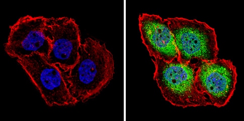
Immunocytochemistry - Anti-PSMB5/MB1 antibody (ab3330)
Immunocytochemistry/Immunofluorescence analysis of PSMB5/MB1 (green) showing staining in the cytoplasm and nucleus of BAEC (Bovine aortic endothelial cell line) cells (right) compared to a negative control without primary antibody (left). Formalin-fixed cells were permeabilized with 0.1% Triton X-100 in TBS for 5-10 minutes and blocked with 3% BSA-PBS for 30 minutes at room temperature. Cells were incubated with ab3330 in 3% BSA-PBS at a dilution of 1:100 and incubated overnight at 4ºC in a humidified chamber. Cells were washed with PBST and incubated with a DyLight-conjugated secondary antibody in PBS at room temperature in the dark. F-actin (red) was stained with a fluorescent red phalloidin and nuclei (blue) were stained with Hoechst or DAPI. Images were taken at a magnification of 60x.

Immunocytochemistry - Anti-PSMB5/MB1 antibody (ab3330)
Immunocytochemistry/Immunofluorescence analysis of PSMB5/MB1 (green) showing staining in the cytoplasm and nucleus of HeLa (Human epithelial adenocarcinoma cell line) cells (right) compared to a negative control without primary antibody (left). Formalin-fixed cells were permeabilized with 0.1% Triton X-100 in TBS for 5-10 minutes and blocked with 3% BSA-PBS for 30 minutes at room temperature. Cells were incubated with ab3330 in 3% BSA-PBS at a dilution of 1:100 and incubated overnight at 4ºC in a humidified chamber. Cells were washed with PBST and incubated with a DyLight-conjugated secondary antibody in PBS at room temperature in the dark. F-actin (red) was stained with a fluorescent red phalloidin and nuclei (blue) were stained with Hoechst or DAPI. Images were taken at a magnification of 60x.


