Anti-Proteasome 26S S3/PSMD3抗体
参阅全部 Proteasome 26S S3/PSMD3 一抗
兔多克隆抗体to Proteasome 26S S3/PSMD3
Rabbit
Detects proteasome 26S subunit S3.
适用于: Flow Cyt, ICC/IFmore details
与反应: Mouse, Human
预测可用于: Cow, Drosophila melanogaster, Non human primates![]()
Synthetic peptide corresponding to Human Proteasome 26S S3/PSMD3 aa 513-534.
Sequence:
EREQQDLEFAKEMAEDDDDSFP
The Life Science industry has been in the grips of a reproducibility crisis for a number of years. Abcam is leading the way in addressing this with our range of recombinant monoclonal antibodies and knockout edited cell lines for gold-standard validation. Please check that this product meets your needs before purchasing.
If you have any questions, special requirements or concerns, please send us an inquiry and/or contact our Support team ahead of purchase. Recommended alternatives for this product can be found below, along with publications, customer reviews and Q&As
Liquid
Shipped at 4°C. Store at +4°C short term (1-2 weeks). Upon delivery aliquot. Store at -20°C or -80°C. Avoid freeze / thaw cycle.
Constituents: 0.1% BSA, 99% PBS
浓度
100 µg 浓度为 1 mg/ml
Immunogen affinity purified
多克隆
IgG
Abpromise™承诺保证使用ab3316于以下的经测试应用
“应用说明”部分 下显示的仅为推荐的起始稀释度;实际最佳的稀释度/浓度应由使用者检定。
| 应用 | Ab评论 | 说明 |
|---|---|---|
| Flow Cyt | Use a concentration of 1 - 20 µg/ml. | |
| ICC/IF | 1/50 - 1/500. |
Entrez Gene: 5709 Human
Entrez Gene: 22123 Mouse
SwissProt: O43242 Human
SwissProt: P14685 Mouse
Unigene: 12970 Human
Unigene: 12194 Mouse
26S proteasome regulatory subunit S3 antibody
OTTHUMP00000164347 antibody
P58 antibody
Proteasome (prosome, macropain) 26S subunit non ATPase 3 antibody
Proteasome subunit p58 antibody
PSMD3 antibody
PSMD3_HUMAN antibody
RPN3 antibody
S3 antibody
Tissue specific transplantation antigen 2 antibody
TSTA2 antibody
26S proteasome non ATPase regulatory subunit 3 antibody
26S proteasome non-ATPase regulatory subunit 3 antibody
26S proteasome regulatory subunit RPN3 antibody
Immunocytochemistry/ Immunofluorescence - Anti-Proteasome 26S S3/PSMD3 antibody (ab3316)
Immunocytochemistry/Immunofluorescence analysis of Proteasome 26S S3/PSMD3 (green) showing staining in the cytoplasm and nucleus of A549 cells (right) compared to a negative control without primary antibody (left). Formalin-fixed cells were permeabilized with 0.1% Triton X-100 in TBS for 5-10 minutes and blocked with 3% BSA-PBS for 30 minutes at room temperature. Cells were incubated with ab3316 in 3% BSA-PBS at a dilution of 1:100 and incubated overnight at 4ºC in a humidified chamber. Cells were washed with PBST and incubated with a DyLight-conjugated secondary antibody in PBS at room temperature in the dark. F-actin (red) was stained with a fluorescent red phalloidin and nuclei (blue) were stained with Hoechst or DAPI. Images were taken at a magnification of 60x.

Flow Cytometry - Anti-Proteasome 26S S3/PSMD3 antibody (ab3316)
Flow Cytometry analysis of HeLa cells labelng Proteasome 26S S3/PSMD3 with ab3316 (Pink) or a rabbit IgG isotype control (Black) 10 µg/mL. Goat anti-Rabbit IgG (H+L) Superclonal™ Alexa Fluor® 647 conjugate at a dilution of 1/50 was used as the Secondary Antibody.

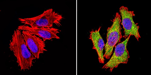
Immunocytochemistry/ Immunofluorescence - Anti-Proteasome 26S S3/PSMD3 antibody (ab3316)
Immunocytochemistry/Immunofluorescence analysis of Proteasome 26S S3/PSMD3 (green) showing staining in the cytoplasm and nucleus of HeLa cells (right) compared to a negative control without primary antibody (left). Formalin-fixed cells were permeabilized with 0.1% Triton X-100 in TBS for 5-10 minutes and blocked with 3% BSA-PBS for 30 minutes at room temperature. Cells were incubated with ab3316 in 3% BSA-PBS at a dilution of 1:100 and incubated overnight at 4ºC in a humidified chamber. Cells were washed with PBST and incubated with a DyLight-conjugated secondary antibody in PBS at room temperature in the dark. F-actin (red) was stained with a fluorescent red phalloidin and nuclei (blue) were stained with Hoechst or DAPI. Images were taken at a magnification of 60x.

Immunocytochemistry/ Immunofluorescence - Anti-Proteasome 26S S3/PSMD3 antibody (ab3316)
Immunocytochemistry/Immunofluorescence analysis of Proteasome 26S S3/PSMD3 (green) showing staining in the cytoplasm and nucleus of NIH-3T3 cells (right) compared to a negative control without primary antibody (left). Formalin-fixed cells were permeabilized with 0.1% Triton X-100 in TBS for 5-10 minutes and blocked with 3% BSA-PBS for 30 minutes at room temperature. Cells were incubated with ab3316 in 3% BSA-PBS at a dilution of 1:100 and incubated overnight at 4ºC in a humidified chamber. Cells were washed with PBST and incubated with a DyLight-conjugated secondary antibody in PBS at room temperature in the dark. F-actin (red) was stained with a fluorescent red phalloidin and nuclei (blue) were stained with Hoechst or DAPI. Images were taken at a magnification of 60x.
抱歉,暂无浏览记录


