Anti-Hsp90 beta抗体
参阅全部 Hsp90 beta 一抗
兔多克隆抗体to Hsp90 beta
Rabbit
Detects heat shock protein 90 beta (HSP90). This antibody does not detect HSP86 alpha.
适用于: ICC/IF, IP, WB, Flow Cyt, IHC-Pmore details
与反应: Mouse, Rat, Human, Non human primates, African green monkey
预测可用于: Rabbit, Horse, Cow, Cynomolgus monkey![]()
Synthetic peptide corresponding to Mouse Hsp90 beta aa 2-13.
Sequence:
PEEVHHGEEEVE
WB: HeLa, MCF7, 293T, K562, A431, HepG2, COS7, NIH3T3 and NRK whole cell lysate. ICC/IF: HepG2, U251, HeLa, NIH3T3 and A2058 cells. Flow Cyt: HeLa cells. IP: HeLa cells. IHC-P: Human tonsil tissue, human placenta tissue, human breast carcinoma tissue.
The Life Science industry has been in the grips of a reproducibility crisis for a number of years. Abcam is leading the way in addressing this with our range of recombinant monoclonal antibodies and knockout edited cell lines for gold-standard validation. Please check that this product meets your needs before purchasing.
If you have any questions, special requirements or concerns, please send us an inquiry and/or contact our Support team ahead of purchase. Recommended alternatives for this product can be found below, along with publications, customer reviews and Q&As
Liquid
Shipped at 4°C. Store at +4°C short term (1-2 weeks). Upon delivery aliquot. Store at -20°C or -80°C. Avoid freeze / thaw cycle.
Preservative: 0.05% Sodium azide
Constituents: 0.1% BSA, 99% PBS
浓度
100 µg 浓度为 1 mg/ml
Immunogen affinity purified
多克隆
IgG
Abpromise™承诺保证使用ab2927于以下的经测试应用
“应用说明”部分 下显示的仅为推荐的起始稀释度;实际最佳的稀释度/浓度应由使用者检定。
| 应用 | Ab评论 | 说明 |
|---|---|---|
| ICC/IF | Use a concentration of 10 - 20 µg/ml. | |
| IP | Use at an assay dependent concentration. 2 μg | |
| WB | 1/1000 - 1/20000. | |
| Flow Cyt | Use a concentration of 1 - 20 µg/ml. | |
| IHC-P | Use a concentration of 10 µg/ml. |
Entrez Gene: 3326 Human
Entrez Gene: 15516 Mouse
Omim: 140572 Human
SwissProt: Q4R4T5 Cynomolgus monkey
SwissProt: Q9GKX8 Horse
SwissProt: P08238 Human
SwissProt: P11499 Mouse
Unigene: 509736 Human
Unigene: 2180 Mouse
Unigene: 98667 Rat
90 kda heat shock protein beta HSP90 beta antibody
D6S182 antibody
FLJ26984 antibody
Heat shock 84 kDa antibody
Heat shock 90kD protein 1, beta antibody
Heat shock 90kDa protein 1 beta antibody
Heat shock protein 90 alpha family class B member 1 antibody
Heat shock protein 90 kDa antibody
Heat shock protein 90kDa alpha (cytosolic) class B member 1 antibody
Heat shock protein 90kDa alpha family class B member 1 antibody
Heat shock protein beta antibody
Heat shock protein HSP 90 beta antibody
Heat shock protein HSP 90-beta antibody
HS90B_HUMAN antibody
HSP 84 antibody
HSP 90 antibody
HSP 90 b antibody
HSP 90b antibody
HSP84 antibody
HSP90 BETA antibody
hsp90ab1 antibody
HSP90B antibody
HSPC2 antibody
HSPCB antibody

Western blot - Anti-Hsp90 beta antibody (ab2927)
All lanes : Anti-Hsp90 beta antibody (ab2927)
Lane 1 : CRISPR targeted HSP90 beta knockout HeLa whole cell lysate
Lane 2 : Wild-type HeLa whole cell lysate

Western blot - Anti-Hsp90 beta antibody (ab2927)
All lanes : Anti-Hsp90 beta antibody (ab2927) at 1/1000 dilution
Lane 1 : MCF7 whole cell lysate
Lane 2 : 293T whole cell lysate
Lane 3 : HeLa whole cell lysate
Lane 4 : K562 whole cell lysate
Lane 5 : A431 whole cell lysate
Lane 6 : HepG2 whole cell lysate
Lane 7 : COS7 whole cell lysate
Lane 8 : NIH3T3 whole cell lysate
Lane 9 : NRK whole cell lysate
Lysates/proteins at 50 µg per lane.
Secondary
All lanes : Goat anti-rabbit IgG-HRP at 1/20000 dilution
Samples were loaded onto a 4-20% Tris-HCl polyacrylamide gel. Proteins were transferred to a PVDF membrane and blocked with 5% BSA/TBST for at least 1 hour. The membrane was then probed with primary antibody (ab2927) overnight at 4°C on a rocking platform, washed in TBS-0.1%Tween 20, and probed with secondary antibody for at least one hour. Chemiluminescent detection was performed using SuperSignal West Pico.

Immunocytochemistry/ Immunofluorescence - Anti-Hsp90 beta antibody (ab2927)
Immunocytochemistry/Immunofluorescence analysis of HSP90 beta shows staining in HepG2 cells. HSP 90 beta staining (green), F-Actin staining with Phalloidin (red) and nuclei with DAPI (blue) is shown. Cells were grown on chamber slides and fixed with formaldehyde prior to staining. Cells were probed without (control) or with or ab2927 at a dilution of 1:100 over night at 4°C, washed with PBS and incubated with a DyLight-488 conjugated goat anti-rabbit secondary antibody. Images were taken at 60X magnification.

Western blot - Anti-Hsp90 beta antibody (ab2927)
Western blot of mouse HSP 86 using ab2927.
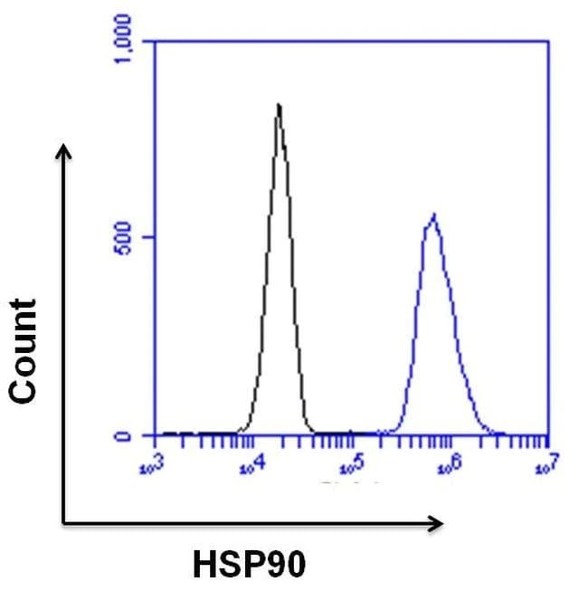
Flow Cytometry - Anti-Hsp90 beta antibody (ab2927)
Flow cytometry analysis of HSP90 was done on HeLa cells. Cells were fixed, permeabilized and stained with a HSP90 rabbit polyclonal antibody (ab2927) (blue histogram) or a rabbit IgG isotype control (black histogram) at a dilution of 10 µg/mL. After incubation for 1 hour on ice, the cells were labeled with a Goat anti-Rabbit IgG (H+L) Superclonal™ Secondary Antibody, Alexa Fluor® 647 conjugate at a dilution of 1/50 for 1 hour on ice. A representative 10,000 cells were acquired and analyzed for each sample.

Immunocytochemistry/ Immunofluorescence - Anti-Hsp90 beta antibody (ab2927)
Immunocytochemistry/Immunofluorescence analysis of HSP90 beta shows staining in U251 cells. HSP 90 beta staining (green), F-Actin staining with Phalloidin (red) and nuclei with DAPI (blue) is shown. Cells were grown on chamber slides and fixed with formaldehyde prior to staining. Cells were probed without (control) or with or ab2927 at a dilution of 1:100 over night at 4°C, washed with PBS and incubated with a DyLight-488 conjugated goat anti-rabbit secondary antibody. Images were taken at 60X magnification.

Immunocytochemistry/ Immunofluorescence - Anti-Hsp90 beta antibody (ab2927)
Immunocytochemistry/Immunofluorescence analysis of HSP90 beta (green) in HeLa and NIH3T3 cells. Formalin fixed cells were permeabilized with 0.1% Triton X-100 in TBS for 10 minutes at room temperature. Cells were blocked with 1% BSA for 15 minutes at room temperature. Cells were incubated with ab2927 at a dilution of 1:50 for at least 1 hour at room temperature, washed with PBS, and incubated with a DyLight 488 goat-anti-rabbit IgG secondary antibody (1:400) for 30 minutes at room temperature. Nuclei (blue) were stained with Hoechst 33342 dye. Images were taken at 20X magnification.

Immunocytochemistry/ Immunofluorescence - Anti-Hsp90 beta antibody (ab2927)
Immunocytochemistry/Immunofluorescence analysis of HSP90 beta shows staining in A2058 cells. HSP 90 beta staining (green), F-Actin staining with Phalloidin (red) and nuclei with DAPI (blue) is shown. Cells were grown on chamber slides and fixed with formaldehyde prior to staining. Cells were probed without (control) or with or ab2927 at a dilution of 1:100 over night at 4°C, washed with PBS and incubated with a DyLight-488 conjugated goat anti-rabbit secondary antibody. Images were taken at 60X magnification.

Immunoprecipitation - Anti-Hsp90 beta antibody (ab2927)
Immunoprecipitation of Hsp90 was performed on HeLa cells. Antigen:antibody complexes were formed by incubating 500µg whole cell lysate with 2µg of Hsp90 polyclonal antibody (ab2927) overnight on a rocking platform at 4°C. Immune complexes were captured on 50µl Protein A/G Agarose washed extensively and eluted with Buffer. Samples were resolved on a 4-20% Tris-HCl polyacrylamide gel, transferred to a PVDF membraneand blocked with 5% BSA/TBST for at least 1 hour. The membrane was probed with a Hsp90 polyclonal antibody (ab2927) at a dilution of 1:1000 overnight rotating at 4°C, washed in TBSTand probed with HRP detection reagent at a dilution of 1:1000 for at least one hour. Chemiluminescent detection was performed.

Immunohistochemistry (Formalin/PFA-fixed paraffin-embedded sections) - Anti-Hsp90 beta antibody (ab2927)
Immunohistochemistry was performed on normal biopsies of deparaffinized Human tonsil tissue. To expose target proteins heat induced antigen retrieval was performed using 10mM sodium citrate (pH6.0) buffer microwaved for 8-15 minutes. Following antigen retrieval tissues were blocked in 3% BSA-PBS for 30 minutes at room temperature. Tissues were then probed at a dilution of 1:100 with a rabbit polyclonal antibody recognizing Heat Shock Protein 84 ab2927 or without primary antibody (negative control) overnight at 4°C in a humidified chamber. Tissues were washed extensively with PBST and endogenous peroxidase activity was quenched with a peroxidase suppressor. Detection was performed using a biotin-conjugated secondary antibody and SA-HRP followed by colorimetric detection using DAB. Tissues were counterstained with hematoxylin and prepped for mounting.

Immunohistochemistry (Formalin/PFA-fixed paraffin-embedded sections) - Anti-Hsp90 beta antibody (ab2927)
Immunohistochemistry was performed on normal biopsies of deparaffinized Human placenta tissue. To expose target proteins heat induced antigen retrieval was performed using 10mM sodium citrate (pH6.0) buffer microwaved for 8-15 minutes. Following antigen retrieval tissues were blocked in 3% BSA-PBS for 30 minutes at room temperature. Tissues were then probed at a dilution of 1:20 with a rabbit polyclonal antibody recognizing Heat Shock Protein 84 ab2927 or without primary antibody (negative control) overnight at 4°C in a humidified chamber. Tissues were washed extensively with PBST and endogenous peroxidase activity was quenched with a peroxidase suppressor. Detection was performed using a biotin-conjugated secondary antibody and SA-HRP followed by colorimetric detection using DAB. Tissues were counterstained with hematoxylin and prepped for mounting.

Immunohistochemistry (Formalin/PFA-fixed paraffin-embedded sections) - Anti-Hsp90 beta antibody (ab2927)
Immunohistochemistry was performed on cancer biopsies of deparaffinized Human breast carcinoma tissue. To expose target proteins heat induced antigen retrieval was performed using 10mM sodium citrate (pH6.0) buffer microwaved for 8-15 minutes. Following antigen retrieval tissues were blocked in 3% BSA-PBS for 30 minutes at room temperature. Tissues were then probed at a dilution of 1:100 with a rabbit polyclonal antibody recognizing Heat Shock Protein 84 ab2927 or without primary antibody (negative control) overnight at 4°C in a humidified chamber. Tissues were washed extensively with PBST and endogenous peroxidase activity was quenched with a peroxidase suppressor. Detection was performed using a biotin-conjugated secondary antibody and SA-HRP followed by colorimetric detection using DAB. Tissues were counterstained with hematoxylin and prepped for mounting.
抱歉,暂无浏览记录


