Anti-Caveolin-3抗体- Caveolae Marker
参阅全部 Caveolin-3 一抗
兔多克隆抗体to Caveolin-3 - Caveolae Marker
Rabbit
This antibody does not detect caveolin-1 or -2.
适用于: IHC-P, ICC/IF, ICC, IHC-Fr, Flow Cyt, WB, IPmore details
与反应: Mouse, Rat, Sheep, Human
Synthetic peptide corresponding to Mouse Caveolin-3 aa 1-19.
Sequence:
MMTEEHTDLEARIIKDIHC
(Peptide available as ab4930)
WB: Rat heart and skeletal muscle; Mouse heart and skeletal muscle; L6 cell lysate. ICC: C2C11 and HeLa cells. IHC-P: Mouse lymph node, heart and skeletal muscle tissues. IP: Mouse heart tissue lysate. Flow Cyt: U-87 MG cells.
The Life Science industry has been in the grips of a reproducibility crisis for a number of years. Abcam is leading the way in addressing this with our range of recombinant monoclonal antibodies and knockout edited cell lines for gold-standard validation. Please check that this product meets your needs before purchasing.
If you have any questions, special requirements or concerns, please send us an inquiry and/or contact our Support team ahead of purchase. Recommended alternatives for this product can be found below, along with publications, customer reviews and Q&As
Liquid
Shipped at 4°C. Store at +4°C short term (1-2 weeks). Upon delivery aliquot. Store at -20°C long term. Avoid freeze / thaw cycle.
Preservative: 0.05% Sodium azide
Constituents: 0.1% BSA, 99% PBS
浓度
100 µg 浓度为 1 mg/ml
Immunogen affinity purified
Antigen affinity chromatography.
多克隆
IgG
Abpromise™承诺保证使用ab2912于以下的经测试应用
“应用说明”部分 下显示的仅为推荐的起始稀释度;实际最佳的稀释度/浓度应由使用者检定。
| 应用 | Ab评论 | 说明 |
|---|---|---|
| IHC-P | (1) | 1/100 - 1/200. |
| ICC/IF | Use at an assay dependent concentration. | |
| ICC | 1/20. | |
| IHC-Fr | (2) | Use at an assay dependent concentration. PubMed: 21408028 |
| Flow Cyt | Use 3-5µg for 106 cells. | |
| WB | (8) | Use a concentration of 1 - 3 µg/ml. |
| IP | Use at an assay dependent concentration. |
Entrez Gene: 859 Human
Entrez Gene: 12391 Mouse
Omim: 601253 Human
SwissProt: P56539 Human
SwissProt: P51637 Mouse
Unigene: 98303 Human
Caveolin-3 antibody
LGMD1C antibody
LQT9 antibody
M-caveolin antibody
MGC126100 antibody
MGC126101 antibody
MGC126129 antibody
OTTHUMP00000115603 antibody
OTTHUMP00000207105 antibody
VIP 21 antibody
VIP21 antibody
CAV3 antibody
CAV3_HUMAN antibody
Caveolin 3 antibody

Western blot - Anti-Caveolin-3 antibody - Caveolae Marker (ab2912)
All lanes : Anti-Caveolin-3 antibody - Caveolae Marker (ab2912)
Lane 1 : Rat heart tissue lysate
Lane 2 : Mouse heart tissue lysate
Lane 3 : HEK293 cell lysate
Lane 4 : Rat skeletal muscle tissue lysate
Lane 5 : Mouse muscle tissue lysate
Lysates/proteins at 20 µg per lane.
Secondary
All lanes : HRP-conjugated goat anti-rabbit IgG (H+L) at 1/2500 dilution
Developed using the ECL technique.
Observed band size: 17 kDawhy is the actual band size different from the predicted?
Blocked with 5% skimmed milk.
Immunocytochemistry - Anti-Caveolin-3 antibody - Caveolae Marker (ab2912)
Immunocytochemistry analysis of Caveolin-3 in HeLa Cells. Cells were grown on chamber slides and fixed with formaldehyde prior to staining. Cells were probed without (control) (right panel) or with ab2912 at a dilution of 1/20 overnight at 4°C, washed with PBS and incubated with a DyLight-488 conjugated secondary antibody. Caveolin-3 staining (green), F-Actin staining with Phalloidin (red) and nuclei with DAPI (blue) is shown. Images were taken at 60X magnification.
Immunohistochemistry (Formalin/PFA-fixed paraffin-embedded sections) - Anti-Caveolin-3 antibody - Caveolae Marker (ab2912)
Immunohistochemistry was performed on normal biopsies of deparaffinized mouse heart tissue. To expose target proteins heat induced antigen retrieval was performed using 10mM sodium citrate (pH6.0) buffer microwaved for 8-15 minutes. Following antigen retrieval tissues were blocked in 3% BSA-PBS for 30 minutes at room temperature. Tissues were then probed at a dilution of 1/200 with a rabbit polyclonal antibody recognizing Caveolin-3 ab2912 or without primary antibody (negative control) overnight at 4°C in a humidified chamber. Tissues were washed extensively with PBST and endogenous peroxidase activity was quenched with a peroxidase suppressor. Detection was performed using a biotin-conjugated secondary antibody and SA-HRP followed by colorimetric detection using DAB. Tissues were counterstained with hematoxylin and prepped for mounting.

Western blot - Anti-Caveolin-3 antibody - Caveolae Marker (ab2912)
All lanes : Anti-Caveolin-3 antibody - Caveolae Marker (ab2912) at 1/1000 dilution
Lane 1 : L6 (Rat skeletal muscle cell line) whole cell lysate
Lane 2 : Mouse skeletal muscle tissue lysate
Lane 3 : Mouse heart tissue lysate
Lane 4 : Mouse brain tissue lysate
Lane 5 : Mouse kidney tissue lysate
Lane 6 : Rat skeletal muscle tissue lysate
Lane 7 : Rat heart tissue lysate
Lane 8 : Rat brain tissue lysate
Lane 9 : Rat kidney tissue lysate
Lysates/proteins at 30 µg per lane.
Observed band size: 17 kDawhy is the actual band size different from the predicted?
Western blot was performed using ab2912 and a ~17 kDa band corresponding to Caveolin 3 was observed. Whole cell extracts (30 µg lysate) were electrophoresed using NuPAGE® 4-12 % Bis-Tris gel. Resolved proteins were then transferred onto a nitrocellulose membrane by iBlot® 2 Dry Blotting System. The blot was probed with the primary antibody (1:1000 dilution) and detected by chemiluminescence with Goat anti-Rabbit IgG (H+L) Superclonal™ Recombinant Secondary Antibody, HRP using the iBright FL 1000. Chemiluminescent detection was performed using Novex® ECL Chemiluminescent Substrate Reagent Kit.
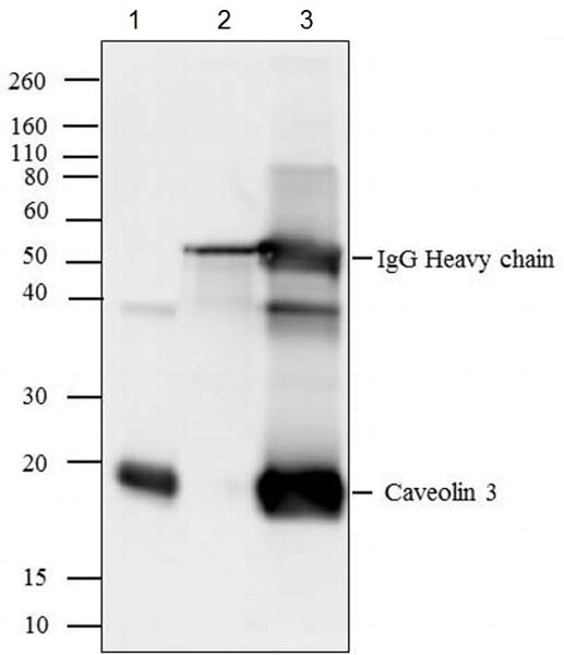
Immunoprecipitation - Anti-Caveolin-3 antibody - Caveolae Marker (ab2912)
Caveolin-3 was immunoprecipitated using 5 µg of ab2912 from mouse heart tissue lysate (Lane 3) using the protein A beads. Normal rabbit IgG was used as a isotype control (Lane 2). 10% input represents the cell extract used for immunoprecipitation (Lane 1). Western blot analysis was performed using ab2912 and HRP-conjugated goat anti-rabbit IgG (H+L) at a dilution of 1/2500. Chemiluminescent detection was performed.

Flow Cytometry - Anti-Caveolin-3 antibody - Caveolae Marker (ab2912)
Flow cytometry analysis of U-87 MG cells. Cells were fixed with 70% ethanol for 10 minutes, permeabilized with 0.25% Triton X-100 for 20 minutes, and blocked with 5% BSA for 30 minutes at room temperature. Cells were labeled with ab2912 (red histogram) or with rabbit isotype control (pink histogram) at 3-5 ug/million cells in 2.5% BSA. After incubation at room temperature for 2 hours, the cells were labeled with Alexa Fluor® 488-conjugated goat anti-rabbit secondary antibody at a dilution of 1/400 for 30 minutes at room temperature. The purple histogram represents unstained control cells and the green histogram represents no-primary-antibody control.

Immunocytochemistry - Anti-Caveolin-3 antibody - Caveolae Marker (ab2912)
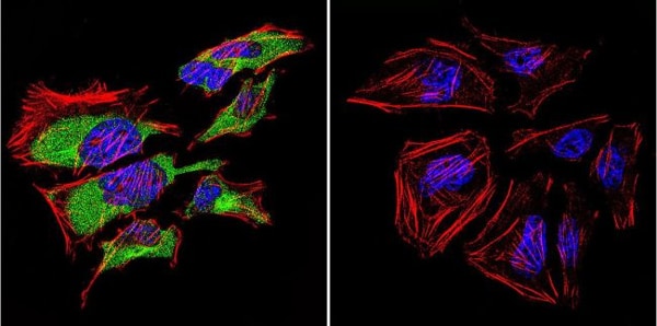
Immunocytochemistry analysis of Caveolin-3 in C2C11 Cells. Cells were grown on chamber slides and fixed with formaldehyde prior to staining. Cells were probed without (control) (right panel) or with ab2912 at a dilution of 1/20 overnight at 4 C, washed with PBS and incubated with a DyLight-488 conjugated secondary antibody. Caveolin-3 staining (green), F-Actin staining with Phalloidin (red) and nuclei with DAPI (blue) is shown. Images were taken at 60X magnification.

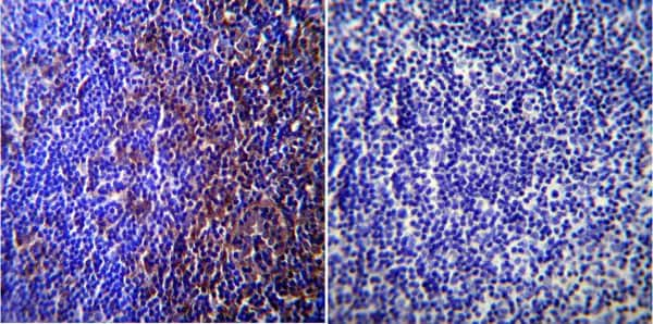
Immunohistochemistry (Formalin/PFA-fixed paraffin-embedded sections) - Anti-Caveolin-3 antibody - Caveolae Marker (ab2912)
Immunohistochemistry was performed on normal biopsies of deparaffinized Mouse skeletal muscle tissue. To expose target proteins heat induced antigen retrieval was performed using 10mM sodium citrate (pH6.0) buffer microwaved for 8-15 minutes. Following antigen retrieval tissues were blocked in 3% BSA-PBS for 30 minutes at room temperature. Tissues were then probed at a dilution of 1/100 with a rabbit polyclonal antibody recognizing Caveolin-3 ab2912 or without primary antibody (negative control) overnight at 4°C in a humidified chamber. Tissues were washed extensively with PBST and endogenous peroxidase activity was quenched with a peroxidase suppressor. Detection was performed using a biotin-conjugated secondary antibody and SA-HRP followed by colorimetric detection using DAB. Tissues were counterstained with hematoxylin and prepped for mounting.

Western blot - Anti-Caveolin-3 antibody - Caveolae Marker (ab2912)
Anti-Caveolin-3 antibody - Caveolae Marker (ab2912) + Rat cardiac muscle tissue lysate
Immunohistochemistry (Formalin/PFA-fixed paraffin-embedded sections) - Anti-Caveolin-3 antibody - Caveolae Marker (ab2912)
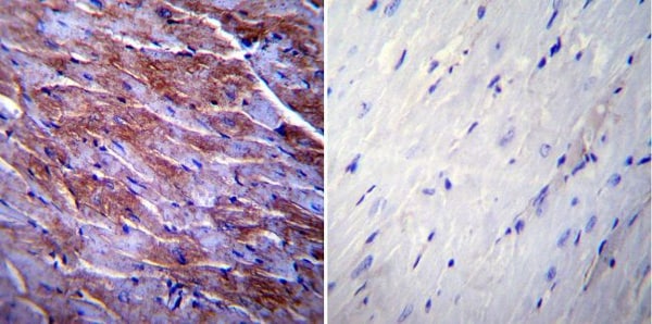
Immunohistochemistry was performed on normal biopsies of deparaffinized mouse lymph node tissue. To expose target proteins heat induced antigen retrieval was performed using 10mM sodium citrate (pH6.0) buffer microwaved for 8-15 minutes. Following antigen retrieval tissues were blocked in 3% BSA-PBS for 30 minutes at room temperature. Tissues were then probed at a dilution of 1/200 with a rabbit polyclonal antibody recognizing Caveolin-3 ab2912 or without primary antibody (negative control) overnight at 4°C in a humidified chamber. Tissues were washed extensively with PBST and endogenous peroxidase activity was quenched with a peroxidase suppressor. Detection was performed using a biotin-conjugated secondary antibody and SA-HRP followed by colorimetric detection using DAB. Tissues were counterstained with hematoxylin and prepped for mounting.

Immunocytochemistry/ Immunofluorescence - Anti-Caveolin-3 antibody - Caveolae Marker (ab2912)
Immunocytochemistry/Immunofluorescence analysis of 70% confluent log phase A-375 cells. Cells were fixed with 4% paraformaldehyde for 15 minutes, permeabilized with 0.25% Triton X-100 for 10 minutes, and blocked with 5% BSA for 1 hour at room temperature. Samples were incubated with ab2912 at 1µg/ml in 1% BSA for 3 hours at room temperature and then labelled with Alexa Fluor® 488-conjugated goat anti-rabbit IgG (H+L) at a dilution of 1/2000 for 45 minutes at room temperature (panel a: green). Nuclei (panel b: blue) were stained with DAPI. F-actin (panel c: red) was stained with Alexa Fluor® 555 Rhodamine Phalloidin (1/300). Panel d is a merged image showing cytoplasmic localization. Panel e is a no primary antibody control. The images were captured at 60X magnification.
抱歉,暂无浏览记录


