Anti-Calreticulin抗体- ER Marker
参阅全部 Calreticulin 一抗
兔多克隆抗体to Calreticulin - ER Marker
Rabbit
适用于: ICC/IF, WBmore details
与反应: Mouse, Rat, Human
Recombinant full length protein corresponding to Human Calreticulin.
WB: HL-60, LNCaP, HeLa and MCF-7 cell lysates; Mouse and rat liver tissue lysates; Mouse skeletal muscle whole cell lysate. ICC/IF: A431, HeLa, U2OS, HepG2 and HMVEC cells.
The Life Science industry has been in the grips of a reproducibility crisis for a number of years. Abcam is leading the way in addressing this with our range of recombinant monoclonal antibodies and knockout edited cell lines for gold-standard validation. Please check that this product meets your needs before purchasing.
If you have any questions, special requirements or concerns, please send us an inquiry and/or contact our Support team ahead of purchase. Recommended alternatives for this product can be found below, along with publications, customer reviews and Q&As
Liquid
Shipped at 4°C. Store at +4°C short term (1-2 weeks). Upon delivery aliquot. Store at -20°C or -80°C. Avoid freeze / thaw cycle.
Preservative: 0.05% Sodium azide
Whole antiserum
多克隆
IgG
Abpromise™承诺保证使用ab2907于以下的经测试应用
“应用说明”部分 下显示的仅为推荐的起始稀释度;实际最佳的稀释度/浓度应由使用者检定。
| 应用 | Ab评论 | 说明 |
|---|---|---|
| ICC/IF | (8) | Use at an assay dependent concentration. |
| WB | (4) | 1/1000. |
Entrez Gene: 811 Human
Entrez Gene: 12317 Mouse
Omim: 109091 Human
SwissProt: P27797 Human
SwissProt: P14211 Mouse
Unigene: 515162 Human
Unigene: 1971 Mouse
Unigene: 467043 Mouse
Unigene: 974 Rat
Autoantigen RO antibody
CALR antibody
CALR protein antibody
CALR_HUMAN antibody
Calregulin antibody
Calreticulin antibody
cC1qR antibody
CRP55 antibody
CRT antibody
CRTC antibody
Endoplasmic reticulum resident protein 60 antibody
Epididymis secretory sperm binding protein Li 99n antibody
ERp60 antibody
FLJ26680 antibody
grp60 antibody
HACBP antibody
HEL S 99n antibody
RO antibody
Sicca syndrome antigen A (autoantigen Ro; calreticulin) antibody
Sicca syndrome antigen A antibody
SSA antibody

Western blot - Anti-Calreticulin antibody - ER Marker (ab2907)
All lanes : Anti-Calreticulin antibody - ER Marker (ab2907) at 1/1000 dilution
Lane 1 : HL-60 cell lysate
Lane 2 : LNCaP cell lysate
Lane 3 : HeLa cell lysate
Lane 4 : MCF-7 cell lysate
Lane 5 : Mouse liver tissue lysate
Lane 6 : Rat liver tissue lysate
Lysates/proteins at 30 µg per lane.
Secondary
All lanes : Goat anti-Rabbit IgG (H+L) Superclonal™ Recombinant Secondary Antibody, HRP at 1/4000 dilution
Observed band size: 55 kDawhy is the actual band size different from the predicted?

Immunocytochemistry/ Immunofluorescence - Anti-Calreticulin antibody - ER Marker (ab2907)Image from Yount JS et al, J Biol Chem. 2012 Jun 1;287(23):19631-41. Epub 2012 Apr 17, Fig 3. DOI 10.1074/jbc.M112.362095 June 1, 2012 The Journal of Biological Chemistry, 287, 19631-19641.
ab2907 used at a 1/1000 dilution staining Calreticulin in HeLa cells by Immunocytochemistry/ Immunofluorescence.HeLa cells were transfected overnight with empty vector or plasmids encoding the indicated IFITM3 constructs. Immunofluorescence with a-HA antibodies allowed IFITM3 visualization, and a-calreticulin staining allowed visualization of the ER. TOPRO-3 was used to visualize nuclei. Scale bars indicate 10 µm. UbΔ indicates mutation of Lys-24, Lys-83, Lys-88, and Lys-104 to alanine.

Immunocytochemistry/ Immunofluorescence - Anti-Calreticulin antibody - ER Marker (ab2907)
Immunocytochemistry/Immunofluorescence analysis of Calreticulin (green) in A431 cells. Formalin fixed cells were permeabilized with 0.1% Triton X-100 in PBS for 10 minutes at room temperature and blocked with 2% BSA in PBS + 0.1% Triton X-100 for 30 minutes at room temperature. Cells were incubated with ab2907 (1:75) for at least 1 hour at room temperature, washed with PBS, and incubated with DyLight 488 goat anti-rabbit IgG secondary antibody (1:250) for 30 minutes at room temperature. Actin was stained with DyLight 650 Phalloidin (1:120) and nuclei (blue) were stained with Hoechst (1ug/ml) for 30 minutes. Images were taken at 20X magnification.

Western blot - Anti-Calreticulin antibody - ER Marker (ab2907)This image is courtesy of an anonymous Abreview
All lanes : Anti-Calreticulin antibody - ER Marker (ab2907) at 1/1000 dilution
All lanes : Whole cell lysate prepared from mouse skeletal muscle
Lysates/proteins at 30 µg per lane.
Secondary
All lanes : HRP-conjugated mouse polyclonal to rabbit Ig at 1/10000 dilution
Developed using the ECL technique.
Performed under reducing conditions.
Exposure time: 3 seconds
Immunocytochemistry/ Immunofluorescence - Anti-Calreticulin antibody - ER Marker (ab2907)
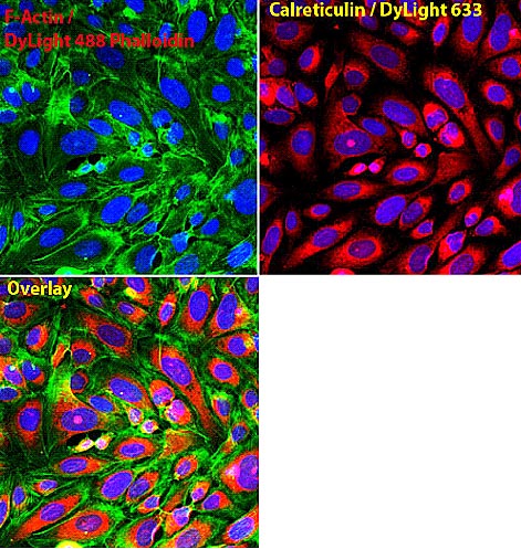
Immunocytochemistry/Immunofluorescence analysis of Calreticulin (red) in U2OS cells. Formalin fixed cells were permeabilized with 0.1% Triton X-100 in PBS for 10 minutes at room temperature and blocked with 2% BSA in PBS + 0.1% Triton X-100 for 30 minutes at room temperature. Cells were incubated with ab2907 (1:75) for at least 1 hour at room temperature, washed with PBS, and incubated with DyLight 633 goat anti-rabbit IgG secondary antibody (1:250) for 30 minutes at room temperature. Actin was stained with DyLight 488 Phalloidin (1:300) and nuclei (blue) were stained with Hoechst (1ug/ml) for 30 minutes. Images were taken at 20X magnification.

Immunocytochemistry/ Immunofluorescence - Anti-Calreticulin antibody - ER Marker (ab2907)
Immunocytochemsitry/Immunofluorescence analysis of Calreticulin (green) U2OS cells. Formalin fixed cells were permeabilized with 0.1% Triton X-100 in PBS for 10 minutes at room temperature and blocked with 2% BSA in PBS 0.1% triton-X for 30 minutes at room temperature. Cells were incubated with ab2907 (1:50) for at least 1 hour at room temperature. Cells were washed with PBS and incubated with DyLight 488 goat-anti-rabbit IgG secondary antibody (1:250) for 30 minutes at room temperature. Actin filaments (red) were stained with DyLight 554-Phalloidin (1:300) in PBS and incubated for 30 minutes. Nuclei (blue) were stained with Hoechst 33342 dye (1µg/mL). Images were taken at 20X magnification.

Immunocytochemistry/ Immunofluorescence - Anti-Calreticulin antibody - ER Marker (ab2907)
Immunocytochemistry/Immunofluorescence analysis of Calreticulin (green) in A431 cells. Formalin fixed cells were permeabilized with 0.1% Triton X-100 in PBS for 10 minutes at room temperature and blocked with 2% BSA in PBS + 0.1% Triton X-100 for 30 minutes at room temperature. Cells were incubated with ab2907 (1:75) for at least 1 hour at room temperature, washed with PBS, and incubated with Dylight 488 goat anti-rabbit IgG secondary antibody (1:250) for 30 minutes at room temperature. Actin was stained with Dylight 350 Phalloidin (1:120) and nuclei (red) were stained with DRAQ5 (1ug/ml) for 30 minutes. Images were taken at 20X magnification.

Immunocytochemistry/ Immunofluorescence - Anti-Calreticulin antibody - ER Marker (ab2907)
Immunocytochemistry/Immunofluorescence analysis of Calreticulin (green) in A431 cells. Formalin fixed cells were permeabilized with 0.1% Triton X-100 in PBS for 10 minutes at room temperature and blocked with 2% BSA in PBS + 0.1% Triton X-100 for 30 minutes at room temperature. Cells were incubated with ab2907 (1:75) for at least 1 hour at room temperature, washed with PBS, and incubated with DyLight 488 goat anti-rabbit IgG secondary antibody (1:250) for 30 minutes at room temperature. Actin was stained with DyLight 550 Phalloidin (1:120) and nuclei (blue) were stained with Hoechst (1ug/ml) for 30 minutes. Images were taken at 20X magnification.

Immunocytochemistry/ Immunofluorescence - Anti-Calreticulin antibody - ER Marker (ab2907)
Immunocytochemistry/Immunofluorescence analysis of HMVEC cells labelling Calreticulin using ab2907.

Immunocytochemistry/ Immunofluorescence - Anti-Calreticulin antibody - ER Marker (ab2907)This image is courtesy of an anonymous Abreview
Immunofluorescence analysis of HepG2 cells, staining Calreticulin with ab2907.
Cells were fixed with paraformaldehyde, permeabilized with 0.1% Saponin and blocked with 10% serum for 1 hour at 20°C. Samples were incubated with primary antibody (1/200 in PBS + 0.1% saponin) for 1 hour at 20°C. An AlexaFluor®647-conjugated donkey anti-rabbit polyclonal IgG (1/400) was used as the secondary antibody.
抱歉,暂无浏览记录


