Anti-gamma H2A.X (phospho S139)抗体
参阅全部 gamma H2A.X 一抗
兔多克隆抗体to gamma H2A.X (phospho S139)
Rabbit
适用于: ICC/IF, WBmore details
与反应: Mouse, Rat, Human
预测可用于: Chimpanzee![]()
This product was produced with the following immunogens:
Synthetic peptide. This information is proprietary to Abcam and/or its suppliers.
(Peptide available as ab15645)
Synthetic peptide. This information is proprietary to Abcam and/or its suppliers.
(Peptide available as ab15645)
ICC/IF: HeLa UV cells. WB : NIH/3T3 (mouse embryonic fibroblast cell line) nuclear lysate (triton enriched), PC-12 (rat adrenal gland pheochromocytoma cell) nuclear lysate (triton enriched).
ab2893 is batch tested in peptide array, western blot and ICC only, although some customers have successfully used this product in IHC and ChIP (see images below). We would recommend ab81299 as an alternative product for use in IHC and ChIP.
The Life Science industry has been in the grips of a reproducibility crisis for a number of years. Abcam is leading the way in addressing this with our range of recombinant monoclonal antibodies and knockout edited cell lines for gold-standard validation. Please check that this product meets your needs before purchasing.
If you have any questions, special requirements or concerns, please send us an inquiry and/or contact our Support team ahead of purchase. Recommended alternatives for this product can be found below, along with publications, customer reviews and Q&As
Liquid
Shipped at 4°C. Store at +4°C short term (1-2 weeks). Upon delivery aliquot. Store at -20°C or -80°C. Avoid freeze / thaw cycle.
pH: 7.40
Preservative: 0.02% Sodium azide
Constituents: 98.98% PBS, 1% BSA
Batches of this product that have a concentration < 1mg/ml may have BSA added as a stabilising agent. If you would like information about the formulation of a specific lot, please contact our scientific support team who will be happy to help.
浓度
批次浓度范围 50 µg 浓度为 0.9 - 1 mg/ml
Immunogen affinity purified
多克隆
IgG
Abpromise™承诺保证使用ab2893于以下的经测试应用
“应用说明”部分 下显示的仅为推荐的起始稀释度;实际最佳的稀释度/浓度应由使用者检定。
| 应用 | Ab评论 | 说明 |
|---|---|---|
| ICC/IF | (7) | Use a concentration of 0.1 µg/ml. |
| WB | (11) | Use a concentration of 1 µg/ml. Detects a band of approximately 17 kDa (predicted molecular weight: 15 kDa). |
Entrez Gene: 3014 Human
Entrez Gene: 15270 Mouse
Omim: 601772 Human
SwissProt: P16104 Human
SwissProt: P27661 Mouse
Unigene: 477879 Human
Unigene: 245931 Mouse
H2A.X antibody
H2a/x antibody
H2AFX antibody
H2AX antibody
H2AX_HUMAN antibody
Histone H2A.X antibody
H2A histone family member X antibody
H2A histone family member X antibody
H2A.FX antibody

Western blot - Anti-gamma H2A.X (phospho S139) antibody (ab2893)
All lanes : Anti-gamma H2A.X (phospho S139) antibody (ab2893) at 1 µg/ml
Lane 1 : NIH/3T3 (mouse embryonic fibroblast cell line) nuclear lysate (triton enriched)
Lane 2 : PC-12 (rat adrenal gland pheochromocytoma cell) nuclear lysate (triton enriched)
Lysates/proteins at 10 µg per lane.
Secondary
All lanes : Goat polyclonal to Rabbit IgG - H&L - Pre-Adsorbed (HRP) at 1/50000 dilution
Predicted band size: 15 kDa
Observed band size: 17 kDawhy is the actual band size different from the predicted?
Exposure time: 16 minutes
Gel type : MES
Blocking buffer : 2% BSA block

Western blot - Anti-gamma H2A.X (phospho S139) antibody (ab2893)
All lanes : Anti-gamma H2A.X (phospho S139) antibody (ab2893) at 1 µg/ml
Lane 1 : NIH 3T3 nuclear lysate (triton enriched)
Lane 2 : PC12 nuclear lysate (triton enriched)
Lysates/proteins at 10 µg per lane.
Secondary
All lanes : Goat polyclonal to Rabbit IgG - H&L - Pre-Adsorbed (HRP) at 1/50000 dilution
Predicted band size: 15 kDa
Observed band size: 17 kDawhy is the actual band size different from the predicted?
Exposure time: 4 minutes
Gel type: MES
Blocking buffer: 2% BSA block
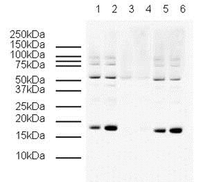
Western blot - Anti-gamma H2A.X (phospho S139) antibody (ab2893)
All lanes : Anti-gamma H2A.X (phospho S139) antibody (ab2893) at 1/500 dilution
Lane 1 : Control HeLa (Human epithelial cell line from cervix adenocarcinoma) whole cell lysate Histone preparation
Lane 2 : Colcemid treated HeLa whole cell lysate Histone preparation
Lane 3 : Control HeLa (Human epithelial cell line from cervix adenocarcinoma) whole cell lysate Histone preparation with Human gamma H2A.X (phospho S139) peptide (ab15645) at 1 µg
Lane 4 : Colcemid treated HeLa whole cell lysate Histone preparation with Human gamma H2A.X (phospho S139) peptide (ab15645) at 1 µg
Lane 5 : Control HeLa (Human epithelial cell line from cervix adenocarcinoma) whole cell lysate Histone preparation with Human Histone H2A.X (unmodified ) peptide (ab15646) at 1 µg
Lane 6 : Colcemid treated HeLa whole cell lysate Histone preparation with Human Histone H2A.X (unmodified ) peptide (ab15646) at 1 µg
Secondary
All lanes : Goat Anti-Rabbit IgG H&L (HRP) (ab6721) at 1/5000 dilution
Predicted band size: 15 kDa
Observed band size: 17 kDawhy is the actual band size different from the predicted?
Additional bands at: 50 kDa (possible cross reactivity)

Immunocytochemistry/ Immunofluorescence - Anti-gamma H2A.X (phospho S139) antibody (ab2893)
ab2893 staining gamma H2A.X (phospho S139) in HeLa UV cells. The cells were fixed with 100% methanol (5 min), permeabilized with 0.1% PBS-Triton X-100 for 5 minutes and then blocked with 1% BSA/10% normal goat serum/0.3M glycine in 0.1%PBS-Tween for 1h. The cells were then incubated overnight at 4°C with ab2893 at 0.1µg/ml and ab7291, Mouse monoclonal [DM1A] to alpha Tubulin - Loading Control. Cells were then incubated with ab150081, Goat polyclonal Secondary Antibody to Rabbit IgG - H&L (Alexa Fluor® 488), pre-adsorbed at 1/1000 dilution (shown in green) and ab150120, Goat polyclonal Secondary Antibody to Mouse IgG - H&L (Alexa Fluor® 594), pre-adsorbed at 1/1000 dilution (shown in pseudocolour red). Nuclear DNA was labelled with DAPI (shown in blue).
Also suitable in cells fixed with 4% paraformaldehyde (10 min).
Image was acquired with a high-content analyser (Operetta CLS, Perkin Elmer) and a maximum intensity projection of confocal sections is shown.

Immunocytochemistry/ Immunofluorescence - Anti-gamma H2A.X (phospho S139) antibody (ab2893)Garcia, C.P. et al PLoS One. 2016; 11(3): e0152607. Fig.2a Published online 2016 Mar 31. doi: 10.1371/journal.pone.0152607 Reproduced under the Creative Commons licence https://creativecommons.org/licenses/by/4.0/
Immunofluorescence photomicrographs of genotoxic-treated (1μM during 3 h) human induced pluripotent stem cells (hiPSCs) and hESCs-derived neuroprogenitors (NP) performed immediately after CPT treatment (1μM during 3 h). The figure shows representative images of cells stained with primary antibodies against ATM phospho-serine1981 (pATM), histoneγH2AX, p53, p53 phospho serine 15 (p53pSer15). Nuclei were counterstained with DAPI. The scale bars represent 100 μm.
Histone gamma H2A.X was detected using ab2893.
From Figure 2a of Garcia et al PLoS One. 2016; 11(3): e0152607. Published online 2016 Mar 31. doi: 10.1371/journal.pone.0152607
Reproduced under the Creative Commons licence: https://creativecommons.org/licenses/by/4.0/

Immunocytochemistry/ Immunofluorescence - Anti-gamma H2A.X (phospho S139) antibody (ab2893)This image is courtesy of Kirk McManus
Asynchronous HeLa cells were paraformaldehyde fixed and immunofluorescently labeled with ab2893 that had been preincubated with either 1) non-phosphorylated or 2) phosphorylated H2AX peptide. Identical exposure times were employed. The Merge images present the DAPI and ab2893 channels as red and green, respectively. Scale bars represent 5µm.
1) Non-phosphorylated peptides
2) Phosphorylated peptides

Western blot - Anti-gamma H2A.X (phospho S139) antibody (ab2893)
HeLa (Human epithelial cell line from cervix adenocarcinoma) cells were incubated at 37°C for 3h with vehicle control (0 µM) and different concentrations of camptothecin (ab120115). Increased expression of γH2A.X (phospho S139) in HeLa cells correlates with an increase in camptothecin concentration, as described in literature.
Whole cell lysates were prepared with RIPA buffer (containing protease inhibitors and sodium orthovanadate), 20µg of each were loaded on the gel and the WB was run under reducing conditions. After transfer the membrane was blocked for an hour using 5% BSA before being incubated with ab2893 at 1 µg/ml and ab10475 at 1 µg/ml overnight at 4°C. Antibody binding was detected using an anti-rabbit antibody conjugated to HRP (ab97051) at 1/10000 dilution and visualised using ECL development solution.

Immunocytochemistry/ Immunofluorescence - Anti-gamma H2A.X (phospho S139) antibody (ab2893)
ab2893 staining γH2A.X in MALME-3M cells treated with terfenadine (ab120270), by ICC/IF. Increase of γH2A.X nuclear expression correlates with increased concentration of terfenadine, as described in literature.
The cells were incubated at 37°C for 6 hours in media containing different concentrations of ab120270 (terfenadine) in DMSO, fixed with 4% formaldehyde for 10 minutes at room temperature and blocked with PBS containing 10% goat serum, 0.3 M glycine, 1% BSA and 0.1% tween for 2h at room temperature. Staining of the treated cells with ab2893 (10 μg/ml) was performed overnight at 4°C in PBS containing 1% BSA and 0.1% tween. A DyLight 488 anti-rabbit polyclonal antibody (ab96899) at 1/250 dilution was used as the secondary antibody. Nuclei were counterstained with DAPI and are shown in blue.

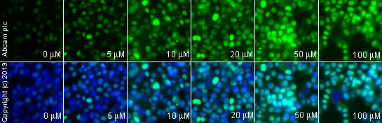
Immunocytochemistry/ Immunofluorescence - Anti-gamma H2A.X (phospho S139) antibody (ab2893)
ab2893 staining γH2AX (phospho S139) in HeLa (Human epithelial cell line from cervix adenocarcinoma) cells treated with camptothecin (ab120115), by ICC/IF. Increased nuclear expression of γH2AX (phospho S139) correlates with increased concentration of camptothecin, as described in literature.
The cells were incubated at 37°C for 3h in media containing different concentrations of ab120115 (camptothecin) in DMSO, fixed with 4% formaldehyde for 10 minutes at room temperature and blocked with PBS containing 10% goat serum, 0.3 M glycine, 1% BSA and 0.1% tween for 2h at room temperature. Staining of the treated cells with ab2893 (10 µg/ml) was performed overnight at 4°C in PBS containing 1% BSA and 0.1% tween. A DyLight 488 goat anti-rabbit polyclonal antibody (ab96899) at 1/250 dilution was used as the secondary antibody. Nuclei were counterstained with DAPI and are shown in blue.

Immunocytochemistry/ Immunofluorescence - Anti-gamma H2A.X (phospho S139) antibody (ab2893)
ab2893 staining γH2A.X in HeLa (Human epithelial cell line from cervix adenocarcinoma) cells treated with SN 38 (ab141108), by ICC/IF. Increase of γH2A.X nuclear expression correlates with increased concentration of SN 38, as described in literature.
The cells were incubated at 37°C for 6 hours in media containing different concentrations of ab141108 (SN 38) in DMSO, fixed with 100% methanol for 5 minutes at -20°C and blocked with PBS containing 10% goat serum, 0.3 M glycine, 1% BSA and 0.1% tween for 2h at room temperature. Staining of the treated cells with ab2893 (5 µg/ml) was performed overnight at 4°C in PBS containing 1% BSA and 0.1% tween. A DyLight 488 anti-rabbit polyclonal antibody (ab96899) at 1/250 dilution was used as the secondary antibody. Nuclei were counterstained with DAPI and are shown in blue.

Immunocytochemistry/ Immunofluorescence - Anti-gamma H2A.X (phospho S139) antibody (ab2893)
ab2893 staining γH2A.X in HeLa (Human epithelial cell line from cervix adenocarcinoma) cells treated with CPT 11 (Irinotecan) (ab141107), by ICC/IF. Increase of γH2A.X nuclear expression correlates with increased concentration of CPT 11 (Irinotecan), as described in literature.
The cells were incubated at 37°C for 6 hours in media containing different concentrations of ab141107 (CPT 11 (Irinotecan)) in DMSO, fixed with 100% methanol for 5 minutes at -20°C and blocked with PBS containing 10% goat serum, 0.3 M glycine, 1% BSA and 0.1% tween for 2h at room temperature. Staining of the treated cells with ab2893 (5 µg/ml) was performed overnight at 4°C in PBS containing 1% BSA and 0.1% tween. A DyLight 488 anti-rabbit polyclonal antibody (ab96899) at 1/250 dilution was used as the secondary antibody. Nuclei were counterstained with DAPI and are shown in blue.

Immunocytochemistry/ Immunofluorescence - Anti-gamma H2A.X (phospho S139) antibody (ab2893)
ab2893 staining γH2A.X in HeLa (Human epithelial cell line from cervix adenocarcinoma) cells treated with 10-Hydroxycamptothecin (ab141071), by ICC/IF. Increase of γH2A.X nuclear expression correlates with increased concentration of 10-Hydroxycamptothecin, as described in literature.
The cells were incubated at 37°C for 6 hours in media containing different concentrations of ab141071 (10-Hydroxycamptothecin) in DMSO, fixed with 100% methanol for 5 minutes at -20°C and blocked with PBS containing 10% goat serum, 0.3 M glycine, 1% BSA and 0.1% tween for 2h at room temperature. Staining of the treated cells with ab2893 (5 µg/ml) was performed overnight at 4°C in PBS containing 1% BSA and 0.1% tween. A DyLight 488 anti-rabbit polyclonal antibody (ab96899) at 1/250 dilution was used as the secondary antibody. Nuclei were counterstained with DAPI and are shown in blue.
Immunocytochemistry/ Immunofluorescence - Anti-gamma H2A.X (phospho S139) antibody (ab2893)
ab2893 staining γH2A.X in weri cells treated with TMPyP4 tosylate (ab120793), by ICC/IF. Increase of γH2A.X nuclear expression correlates with increased concentration of TMPyP4 tosylate, as described in literature.
The cells were incubated at 37°C for 24 hours in media containing different concentrations of ab120793 (TMPyP4 tosylate ) in DMSO, fixed with 4% formaldehyde for 10 minutes at room temperature and blocked with PBS containing 10% goat serum, 0.3 M glycine, 1% BSA and 0.1% tween for 2h at room temperature. Staining of the treated cells with ab2893 (1 μg/ml) was performed overnight at 4°C in PBS containing 1% BSA and 0.1% tween. A DyLight 488 anti-rabbit polyclonal antibody (ab96899) at 1/250 dilution was used as the secondary antibody. Nuclei were counterstained with DAPI and are shown in blue.

Immunocytochemistry/ Immunofluorescence - Anti-gamma H2A.X (phospho S139) antibody (ab2893)Image courtesy of Dr. Kirk McManus
Asynchronous HeLa (Human epithelial cell line from cervix adenocarcinoma) cells were exposed to 2Gy and permitted to recover for 30min. Cells were paraformaldehyde fixed (4%), immunofluorescently labeled with ab2893 and counterstained with DAPI. The merge image presents the DAPI and ab2893 channels as red and green, respectively. The scale bar represents 5µm.
抱歉,暂无浏览记录


