Anti-eIF4G1抗体
参阅全部 eIF4G1 一抗
兔多克隆抗体to eIF4G1
Rabbit
适用于: IP, WB, IHC-P, ICC/IFmore details
与反应: Rat, Human, African green monkey
预测可用于: Mouse, Rabbit, Horse, Hamster, Cow, Cat, Dog, Chimpanzee, Rhesus monkey, Gorilla, Chinese hamster, Orangutan, Elephant![]()
Synthetic peptide within Human eIF4G1 aa 550-650. The exact immunogen sequence used to generate this antibody is proprietary information. If additional detail on the immunogen is needed to determine the suitability of the antibody for your needs, please contact our Scientific Support team to discuss your requirements. NP_886553.2 (GeneID 1981).
Database link: Q04637
WB: HeLa and HEK-293T whole cell lysate. Rat liver lysate. IHC-P: Human colon tissue. IP: eIF4G1 in HeLa whole cell lysate. ICC/IF: MCF7 cells.
The Life Science industry has been in the grips of a reproducibility crisis for a number of years. Abcam is leading the way in addressing this with our range of recombinant monoclonal antibodies and knockout edited cell lines for gold-standard validation. Please check that this product meets your needs before purchasing.
If you have any questions, special requirements or concerns, please send us an inquiry and/or contact our Support team ahead of purchase. Recommended alternatives for this product can be found below, along with publications, customer reviews and Q&As
Liquid
Shipped at 4°C. Upon delivery aliquot and store at -20°C. Avoid freeze / thaw cycles.
pH: 7
Preservative: 0.1% Sodium azide
浓度
50 µg 浓度为 1 mg/ml
Affinity purified using the immunising peptide immobilized on solid support.
多克隆
IgG
Abpromise™承诺保证使用ab2609于以下的经测试应用
“应用说明”部分 下显示的仅为推荐的起始稀释度;实际最佳的稀释度/浓度应由使用者检定。
| 应用 | Ab评论 | 说明 |
|---|---|---|
| IP | (1) | 1/1000. |
| WB | (2) | 1/1000 - 1/10000. Detects a band of approximately 200 kDa (predicted molecular weight: 220 kDa). EIF4G is more susceptible to degradation compared to other proteins, especially from some tissue sources such as liver. This is true even when tissue is stored at frozen. SDS-PAGE sample buffer may improve the stability, but samples that are stored frozen may show degradation bands that have been described in the literature.Therefore if multiple bands are observed in WB, it is likely due to the degradation of eIF4G rather than non-specificity of the antibody. Read More |
| IHC-P | Use a concentration of 4 µg/ml. | |
| ICC/IF | 1/500. |
Entrez Gene: 1981 Human
Entrez Gene: 208643 Mouse
Omim: 600495 Human
SwissProt: Q04637 Human
SwissProt: Q6NZJ6 Mouse
SwissProt: P41110 Rabbit
Unigene: 433750 Human
Unigene: 260256 Mouse
DKFZp686A1451 antibody
eIF 4 gamma 1 antibody
eIF 4G 1 antibody
eIF 4G1 antibody
eIF-4-gamma 1 antibody
eIF-4G 1 antibody
eIF-4G1 antibody
EIF4 gamma antibody
EIF4F antibody
EIF4G antibody
EIF4G1 antibody
EIF4GI antibody
Eukaryotic translation initiation factor 4 gamma 1 antibody
IF4G1_HUMAN antibody
p220 antibody

Western blot - Anti-eIF4G1 antibody (ab2609)
All lanes : Anti-eIF4G1 antibody (ab2609) at 0.1 µg/ml
Lane 1 : HeLa (Human epithelial cell line from cervix adenocarcinoma) whole cell lysate
Lane 2 : HEK-293T (Human epithelial cell line from embryonic kidney transformed with large T antigen) whole cell lysate
Lysates/proteins at 50 µg per lane.
Predicted band size: 220 kDa
Exposure time: 3 minutes
Lysates prepared using NETN lysis buffer.
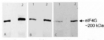
Western blot - Anti-eIF4G1 antibody (ab2609)
50µg (lane 1) and 150µg (lane 2) rat liver lysate, separated on 7.5% acrylamide SDS-PAGE gel. Detected using ab2609 at 1:1000 (A), 1:5000 (B) and 1:10000 (C) dilution by ECL.
50µg (lane 1) and 150µg (lane 2) rat liver lysate, separated on 7.5% acrylamide SDS-PAGE gel. Detected using ab2609 at 1:1000 (A), 1:5000 (B) and 1:10000 (C) dilution by ECL.

Immunohistochemistry (Formalin/PFA-fixed paraffin-embedded sections) - Anti-eIF4G1 antibody (ab2609)
ab2609 (4µg/ml) staining eIF4G1 in human colon using an automated system (DAKO Autostainer Plus). Using this protocol there is strong staining of the cytoplasm of the intestinal cells.
Sections were rehydrated and antigen retrieved with the Dako 3 in 1 AR buffer EDTA pH 9.0 in a DAKO PT link. Slides were peroxidase blocked in 3% H2O2 in methanol for 10 mins. They were then blocked with Dako Protein block for 10 minutes (containing casein 0.25% in PBS) then incubated with primary antibody for 20 min and detected with Dako envision flex amplification kit for 30 minutes. Colorimetric detection was completed with Diaminobenzidine for 5 minutes. Slides were counterstained with Haematoxylin and coverslipped under DePeX. Please note that, for manual staining, optimization of primary antibody concentration and incubation time is recommended. Signal amplification may be required.

Immunoprecipitation - Anti-eIF4G1 antibody (ab2609)
Lane 1: immunoprecipitated by ab2609 at 6 µg per reaction;
Lane 2: Immunoprecipitated by control IgG at 6 µg per reaction.
All lanes : Anti-eIF4G1 antibody (ab2609) at 1 µg/ml
Lane 1 : HeLa (Human epithelial cell line from cervix adenocarcinoma) whole cell lysate
Lane 2 : HeLa whole cell lysate
Exposure time: 10 seconds

Immunocytochemistry/ Immunofluorescence - Anti-eIF4G1 antibody (ab2609)
ICC/IF image of ab2609 stained MCF7 cells. The cells were 4% PFA fixed (10 min) and then incubated in 1%BSA / 10% normal goat serum / 0.3M glycine in 0.1% PBS-Tween for 1h to permeabilise the cells and block non-specific protein-protein interactions. The cells were then incubated with the antibody (ab2609, 1µg/ml) overnight at +4°C. The secondary antibody (green) was Alexa Fluor® 488 goat anti-rabbit IgG (H+L) used at a 1/1000 dilution for 1h. Alexa Fluor® 594 WGA was used to label plasma membranes (red) at a 1/200 dilution for 1h. DAPI was used to stain the cell nuclei (blue) at a concentration of 1.43µM.


